ムービー
ムービー コントローラー


+ データを開く

- 基本情報
基本情報
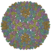
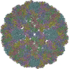
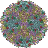
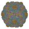
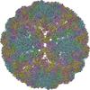
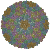
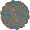
| 登録情報 | データベース: PDB / ID: 1z8y | ||||||
|---|---|---|---|---|---|---|---|
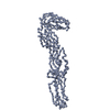
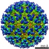
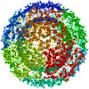
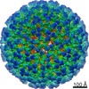
| タイトル | Mapping the E2 Glycoprotein of Alphaviruses | ||||||
 要素 要素 |
| ||||||
 キーワード キーワード | VIRUS / icosahedral enveloped virus / Icosahedral virus | ||||||
| 機能・相同性 |  機能・相同性情報 機能・相同性情報icosahedral viral capsid, spike / togavirin / T=4 icosahedral viral capsid / ubiquitin-like protein ligase binding / symbiont-mediated suppression of host toll-like receptor signaling pathway / clathrin-dependent endocytosis of virus by host cell / host cell cytoplasm / membrane => GO:0016020 / membrane fusion / serine-type endopeptidase activity ...icosahedral viral capsid, spike / togavirin / T=4 icosahedral viral capsid / ubiquitin-like protein ligase binding / symbiont-mediated suppression of host toll-like receptor signaling pathway / clathrin-dependent endocytosis of virus by host cell / host cell cytoplasm / membrane => GO:0016020 / membrane fusion / serine-type endopeptidase activity / fusion of virus membrane with host endosome membrane / viral envelope / host cell nucleus / virion attachment to host cell / host cell plasma membrane / structural molecule activity / virion membrane / proteolysis / RNA binding / membrane 類似検索 - 分子機能 | ||||||
| 生物種 |  Sindbis virus (シンドビスウイルス) Sindbis virus (シンドビスウイルス) | ||||||
| 手法 | 電子顕微鏡法 / 単粒子再構成法 / クライオ電子顕微鏡法 / 解像度: 9 Å | ||||||
 データ登録者 データ登録者 | Mukhopadhyay, S. / Zhang, W. / Gabler, S. / Chipman, P.R. / Strauss, E.G. / Strauss, J.H. / Baker, T.S. / Kuhn, R.J. / Rossmann, M.G. | ||||||
 引用 引用 | ジャーナル: Structure / 年: 2006 タイトル: Mapping the structure and function of the E1 and E2 glycoproteins in alphaviruses. 著者: Suchetana Mukhopadhyay / Wei Zhang / Stefan Gabler / Paul R Chipman / Ellen G Strauss / James H Strauss / Timothy S Baker / Richard J Kuhn / Michael G Rossmann /  要旨: The 9 A resolution cryo-electron microscopy map of Sindbis virus presented here provides structural information on the polypeptide topology of the E2 protein, on the interactions between the E1 and ...The 9 A resolution cryo-electron microscopy map of Sindbis virus presented here provides structural information on the polypeptide topology of the E2 protein, on the interactions between the E1 and E2 glycoproteins in the formation of a heterodimer, on the difference in conformation of the two types of trimeric spikes, on the interaction between the transmembrane helices of the E1 and E2 proteins, and on the conformational changes that occur when fusing with a host cell. The positions of various markers on the E2 protein established the approximate topology of the E2 structure. The largest conformational differences between the icosahedral surface spikes at icosahedral 3-fold and quasi-3-fold positions are associated with the monomers closest to the 5-fold axes. The long E2 monomers, containing the cell receptor recognition motif at their extremities, are shown to rotate by about 180 degrees and to move away from the center of the spikes during fusion. | ||||||
| 履歴 |
| ||||||
| Remark 999 | SEQUENCE Author states that it appears even though the correct sequence utilizes a lysine, leucine ...SEQUENCE Author states that it appears even though the correct sequence utilizes a lysine, leucine was used in the model. |
- 構造の表示
構造の表示
| ムービー |
ムービービューア |
|---|---|
| 構造ビューア | 分子: MolmilJmol/JSmol |
- ダウンロードとリンク
-ダウンロード
| PDBx/mmCIF形式 | 1z8y.cif.gz | 435 KB | 表示 | PDBx/mmCIF形式 |
|---|---|---|---|---|
| PDB形式 | pdb1z8y.ent.gz | 369.2 KB | 表示 | PDB形式 |
| PDBx/mmJSON形式 | 1z8y.json.gz | ツリー表示 | PDBx/mmJSON形式 | |
| その他 |  その他のダウンロード その他のダウンロード |
-検証レポート
| 文書・要旨 | 1z8y_validation.pdf.gz | 1018.5 KB | 表示 | wwPDB検証レポート |
|---|---|---|---|---|
| 文書・詳細版 | 1z8y_full_validation.pdf.gz | 1.2 MB | 表示 | |
| XML形式データ | 1z8y_validation.xml.gz | 90.4 KB | 表示 | |
| CIF形式データ | 1z8y_validation.cif.gz | 127.2 KB | 表示 | |
| アーカイブディレクトリ | https://data.pdbj.org/pub/pdb/validation_reports/z8/1z8yftp://data.pdbj.org/pub/pdb/validation_reports/z8/1z8y | HTTPS FTP |
-関連構造データ
-リンク
 PDBj
PDBj


- 集合体
集合体
| 登録構造単位 | 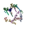
|
|---|---|
| 1 | x 60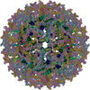
|
| 2 |
|
| 3 | x 5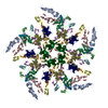
|
| 4 | x 6
|
| 5 | 
|
| 対称性 | 点対称性: (ヘルマン・モーガン記号: 532 / シェーンフリース記号: I (正20面体型対称)) |
-要素
| #1: タンパク質 | 分子量: 31440.787 Da / 分子数: 4 / 断片: E1 ectodomain domains I+II, residues 1-290 / 由来タイプ: 天然 由来: (天然) Sindbis virus (シンドビスウイルス)属: Alphavirus / Cell: Baby Hamster Kidney / 株: TE12 / 参照: UniProt: P03316 #2: タンパク質 | 分子量: 9447.606 Da / 分子数: 4 / 断片: E1 ectodomain domain III, residues 295-383 / 由来タイプ: 天然 由来: (天然) Sindbis virus (シンドビスウイルス)属: Alphavirus / Cell: Baby Hamster Kidney / 株: TE12 / 参照: UniProt: P03316 #3: タンパク質・ペプチド | 分子量: 3410.192 Da / 分子数: 4 / 断片: E1 transmembrane region, residues 409-439 / 由来タイプ: 天然 由来: (天然) Sindbis virus (シンドビスウイルス)属: Alphavirus / Cell: Baby Hamster Kidney / 株: TE12 / 参照: UniProt: P03316 #4: タンパク質・ペプチド | 分子量: 3751.593 Da / 分子数: 4 / 断片: E2 transmembrane region, residues 363-398 / 由来タイプ: 天然 由来: (天然) Sindbis virus (シンドビスウイルス)属: Alphavirus / Cell: Baby Hamster Kidney / 株: TE12 / 参照: UniProt: P11259, UniProt: P03316*PLUS #5: タンパク質 | 分子量: 16545.631 Da / 分子数: 4 / 断片: Capsid protein, residues 114-264 / 由来タイプ: 天然 由来: (天然) Sindbis virus (シンドビスウイルス)属: Alphavirus / Cell: Baby Hamster Kidney / 株: TE12 参照: UniProt: P03316, 加水分解酵素; プロテアーゼ; ペプチド結合加水分解酵素; セリンエンドペプチターゼ |
|---|
-実験情報
-実験
| 実験 | 手法: 電子顕微鏡法 |
|---|---|
| EM実験 | 試料の集合状態: PARTICLE / 3次元再構成法: 単粒子再構成法 |
- 試料調製
試料調製
| 構成要素 |
| |||||||||||||||||||||||||||||||||||
|---|---|---|---|---|---|---|---|---|---|---|---|---|---|---|---|---|---|---|---|---|---|---|---|---|---|---|---|---|---|---|---|---|---|---|---|---|
| ウイルスについての詳細 | ホストのカテゴリ: EUKARYOTES / タイプ: VIRION | |||||||||||||||||||||||||||||||||||
| 天然宿主 | 株: Baby Hamster Kidney | |||||||||||||||||||||||||||||||||||
| 緩衝液 | 名称: 50 mM Tris-Cl, 200 mM NaCl, 0.1 mM EDTA / pH: 7.4 / 詳細: 50 mM Tris-Cl, 200 mM NaCl, 0.1 mM EDTA | |||||||||||||||||||||||||||||||||||
| 試料 | 包埋: NO / シャドウイング: NO / 染色: NO / 凍結: YES | |||||||||||||||||||||||||||||||||||
| 試料支持 | 詳細: See Pletnev et al. (2001) Cell 105:127-136 for experimental details on sample preparation | |||||||||||||||||||||||||||||||||||
| 急速凍結 | 装置: HOMEMADE PLUNGER / 凍結剤: ETHANE |
- 電子顕微鏡撮影
| 顕微鏡 | モデル: FEI/PHILIPS CM200FEG / 日付: 2000年6月21日 |
|---|---|
| 電子銃 | 電子線源:  FIELD EMISSION GUN / 加速電圧: 200 kV / 照射モード: FLOOD BEAM FIELD EMISSION GUN / 加速電圧: 200 kV / 照射モード: FLOOD BEAM |
| 電子レンズ | モード: BRIGHT FIELD / 倍率(公称値): 38000 X / 最大 デフォーカス(公称値): 2580 nm / 最小 デフォーカス(公称値): 1100 nm / Cs: 2 mm |
| 試料ホルダ | 温度: 93.15 K / 傾斜角・最大: 0 ° / 傾斜角・最小: 0 ° |
| 撮影 | 電子線照射量: 18 e/Å2 / フィルム・検出器のモデル: KODAK SO-163 FILM 詳細: THE COORDINATES IN THIS ENTRY WERE GENERATED FROM ELECTRON MICROSCOPY DATA. |
- 解析
解析
| EMソフトウェア |
| ||||||||||||
|---|---|---|---|---|---|---|---|---|---|---|---|---|---|
| CTF補正 | 詳細: Fourier transform of each image was modified | ||||||||||||
| 対称性 | 点対称性: I (正20面体型対称) | ||||||||||||
| 3次元再構成 | 手法: cross-common lines / 解像度: 9 Å / 粒子像の数: 7085 / ピクセルサイズ(実測値): 1.785 Å / 対称性のタイプ: POINT | ||||||||||||
| 原子モデル構築 | プロトコル: RIGID BODY FIT / 空間: REAL / 詳細: REFINEMENT PROTOCOL--rigid body | ||||||||||||
| 原子モデル構築 |
| ||||||||||||
| 精密化ステップ | サイクル: LAST
|