 Movie
Movie Controller


+ Open data

- Basic information
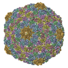
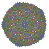
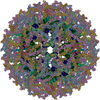
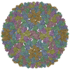
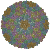
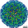
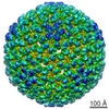
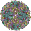
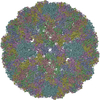
| Entry | Database: PDB / ID: 3ddx | ||||||
|---|---|---|---|---|---|---|---|
| Title | HK97 bacteriophage capsid Expansion Intermediate-II model | ||||||
 Components Components | Major capsid protein | ||||||
 Keywords Keywords | VIRUS / Bacteriophage / HK97 / Capsid Protein / Expansion Intermediate / Virion / icosahedral virus | ||||||
| Function / homology | Phage capsid / Phage capsid family / viral procapsid maturation / T=7 icosahedral viral capsid / viral capsid / identical protein binding / Major capsid protein Function and homology information Function and homology information | ||||||
| Biological species |  Bacteriophage HK97 (virus) Bacteriophage HK97 (virus) | ||||||
| Method | ELECTRON MICROSCOPY / single particle reconstruction / cryo EM | ||||||
 Authors Authors | Lee, K.K. / Gan, L. / Conway, J.F. / Hendrix, R.W. / Steven, A.C. / Johnson, J.E. | ||||||
 Citation Citation | Journal: Structure / Year: 2008 Title: Virus capsid expansion driven by the capture of mobile surface loops. Authors: Kelly K Lee / Lu Gan / Hiro Tsuruta / Crystal Moyer / James F Conway / Robert L Duda / Roger W Hendrix / Alasdair C Steven / John E Johnson /  Abstract: The capsids of tailed-DNA bacteriophages first assemble as procapsids, which mature by converting into a new form that is strong enough to contain a densely packed viral chromosome. We demonstrate ...The capsids of tailed-DNA bacteriophages first assemble as procapsids, which mature by converting into a new form that is strong enough to contain a densely packed viral chromosome. We demonstrate that the intersubunit crosslinking that occurs during maturation of HK97 capsids actually promotes the structural transformation. Small-angle X-ray scattering and crosslinking assays reveal that a shift in the crosslink pattern accompanies conversion of a semimature particle, Expansion Intermediate-I/II, to a more mature state, Balloon. This transition occurs in a switch-like fashion. We find that crosslink formation shifts the global conformational balance to favor the balloon state. A pseudoatomic model of EI-I/II derived from cryo-EM provides insight into the relationship between crosslink formation and conformational switching. | ||||||
| History |
|
- Structure visualization
| Movie |
Movie viewer |
|---|---|
| Structure viewer | Molecule: MolmilJmol/JSmol |
- Downloads & links
-Download
| PDBx/mmCIF format | 3ddx.cif.gz | 266.7 KB | Display | PDBx/mmCIF format |
|---|---|---|---|---|
| PDB format | pdb3ddx.ent.gz | 170.1 KB | Display | PDB format |
| PDBx/mmJSON format | 3ddx.json.gz | Tree view | PDBx/mmJSON format | |
| Others |  Other downloads Other downloads |
-Validation report
| Summary document | 3ddx_validation.pdf.gz | 374.5 KB | Display | wwPDB validaton report |
|---|---|---|---|---|
| Full document | 3ddx_full_validation.pdf.gz | 416.6 KB | Display | |
| Data in XML | 3ddx_validation.xml.gz | 37.5 KB | Display | |
| Data in CIF | 3ddx_validation.cif.gz | 57.1 KB | Display | |
| Arichive directory | https://data.pdbj.org/pub/pdb/validation_reports/dd/3ddxftp://data.pdbj.org/pub/pdb/validation_reports/dd/3ddx | HTTPS FTP |
-Related structure data
| Related structure data | |
|---|---|
| Similar structure data |


-Links
 PDBj
PDBj

- Assembly
| Deposited unit | 
|
|---|---|
| 1 | x 60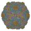
|
| 2 |
|
| 3 | x 5
|
| 4 | x 6
|
| 5 | 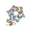
|
| Symmetry | Point symmetry: (Schoenflies symbol: I (icosahedral)) |
-Components
| #1: Protein | Mass: 30804.607 Da / Num. of mol.: 7 Source method: isolated from a genetically manipulated source Source: (gene. exp.) Bacteriophage HK97 (virus) / Gene: 5 / Plasmid: PT7-HD2.9 / Production host:  |
|---|
-Experimental details
-Experiment
| Experiment | Method: ELECTRON MICROSCOPY |
|---|---|
| EM experiment | Aggregation state: PARTICLE / 3D reconstruction method: single particle reconstruction |
- Sample preparation
| Component | Name: Bacteriophage HK97 Expansion Intermediate II / Type: VIRUS |
|---|---|
| Buffer solution | Name: 50mM Na-Acetate, 200mM KCl / pH: 4.18 / Details: 50mM Na-Acetate, 200mM KCl |
| Specimen | Embedding applied: NO / Shadowing applied: NO / Staining applied: NO / Vitrification applied: YES |
- Electron microscopy imaging
| Microscopy | Model: FEI/PHILIPS CM200FEG |
|---|---|
| Electron gun | Electron source:  FIELD EMISSION GUN / Accelerating voltage: 120 kV / Illumination mode: FLOOD BEAM FIELD EMISSION GUN / Accelerating voltage: 120 kV / Illumination mode: FLOOD BEAM |
| Electron lens | Mode: BRIGHT FIELD / Nominal magnification: 38000 X |
- Processing
| EM software |
| ||||||||||||
|---|---|---|---|---|---|---|---|---|---|---|---|---|---|
| Symmetry | Point symmetry: I (icosahedral) | ||||||||||||
| 3D reconstruction | Symmetry type: POINT | ||||||||||||
| Atomic model building | Protocol: RIGID BODY FIT / Space: RECIPROCAL / Target criteria: Vector Reciprocal Space Target Details: METHOD--manual and rigid body fitting. THE COORDINATES CONTAIN ONLY THE BACKBONE ATOMS. DURING RIGID BODY DOCKING OF THE PROTEIN MODEL INTO THE EM DENSITY, THE TWO SUBDOMAINS 104-125 AND 155- ...Details: METHOD--manual and rigid body fitting. THE COORDINATES CONTAIN ONLY THE BACKBONE ATOMS. DURING RIGID BODY DOCKING OF THE PROTEIN MODEL INTO THE EM DENSITY, THE TWO SUBDOMAINS 104-125 AND 155-175 OF EACH SUBUNIT WERE ALLOWED TO MOVE INDEPENDENTLY FROM THE REMAINDER OF THE RESIDUES (THE CORE OF EACH SUBUNIT). THAT IS WHY THE CONNECTIVITY OF THOSE SEGMENTS WITH THE REST OF THE SUBUNIT IS NOT WELL-PRESERVED. REFINEMENT PROTOCOL--Rigid Body, Magnification Optimized using Van Der Waals Constraints and Real-space Correlation Coefficient | ||||||||||||
| Atomic model building | PDB-ID: 1OHG Accession code: 1OHG / Source name: PDB / Type: experimental model | ||||||||||||
| Refinement step | Cycle: LAST
|
