 ムービー
ムービー コントローラー


+ データを開く

- 基本情報
基本情報
| 登録情報 | データベース: PDB / ID: 1gw7 | ||||||
|---|---|---|---|---|---|---|---|
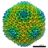
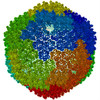
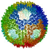
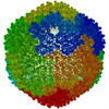
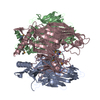
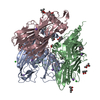
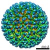
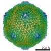
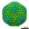
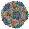
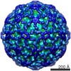
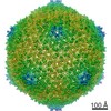
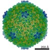
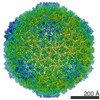
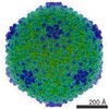
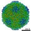
| タイトル | QUASI-ATOMIC RESOLUTION MODEL OF BACTERIOPHAGE PRD1 CAPSID, OBTAINED BY COMBINED CRYO-EM AND X-RAY CRYSTALLOGRAPHY. | ||||||
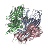
 要素 要素 | MAJOR CAPSID PROTEIN | ||||||
 キーワード キーワード | VIRUS/VIRAL PROTEIN /  TECTIVIRIDAE / BACTERIOPHAGE PRD1 / CRYO- EM / IMAGE RECONSTRUCTION / ICOSAHEDRAL VIRUS / VIRUS-VIRAL PROTEIN complex TECTIVIRIDAE / BACTERIOPHAGE PRD1 / CRYO- EM / IMAGE RECONSTRUCTION / ICOSAHEDRAL VIRUS / VIRUS-VIRAL PROTEIN complex | ||||||
| 機能・相同性 | Bacteriophage PRD1, P3 / Bacteriophage PRD1, P3, N-terminal / P3 major capsid protein / Group II dsDNA virus coat/capsid protein / Viral coat protein subunit / カプシド / Major capsid protein P3 機能・相同性情報 機能・相同性情報 | ||||||
| 生物種 |   BACTERIOPHAGE PRD1 (ファージ) BACTERIOPHAGE PRD1 (ファージ) | ||||||
| 手法 | 電子顕微鏡法 / 単粒子再構成法 / クライオ電子顕微鏡法 / 解像度: 13.5 Å | ||||||
 データ登録者 データ登録者 | San Martin, C. / Huiskonen, J. / Bamford, J.K.H. / Butcher, S.J. / Fuller, S.D. / Bamford, D.H. / Burnett, R.M. | ||||||
 引用 引用 | ジャーナル: Nat Struct Biol / 年: 2002 タイトル: Minor proteins, mobile arms and membrane-capsid interactions in the bacteriophage PRD1 capsid. 著者: Carmen San Martín / Juha T Huiskonen / Jaana K H Bamford / Sarah J Butcher / Stephen D Fuller / Dennis H Bamford / Roger M Burnett /  要旨: Bacteriophage PRD1 shares many structural and functional similarities with adenovirus. A major difference is the PRD1 internal membrane, which acts in concert with vertex proteins to translocate the ...Bacteriophage PRD1 shares many structural and functional similarities with adenovirus. A major difference is the PRD1 internal membrane, which acts in concert with vertex proteins to translocate the phage genome into the host. Multiresolution models of the PRD1 capsid, together with genetic analyses, provide fine details of the molecular interactions associated with particle stability and membrane dynamics. The N- and C-termini of the major coat protein (P3), which are required for capsid assembly, act as conformational switches bridging capsid to membrane and linking P3 trimers. Electrostatic P3-membrane interactions increase virion stability upon DNA packaging. Newly revealed proteins suggest how the metastable vertex works and how the capsid edges are stabilized. #1: ジャーナル: Acta Crystallogr D Biol Crystallogr / 年: 2002タイトル: The X-ray crystal structure of P3, the major coat protein of the lipid-containing bacteriophage PRD1, at 1.65 A resolution. 著者: Stacy D Benson / Jaana K H Bamford / Dennis H Bamford / Roger M Burnett / 要旨: P3 has been imaged with X-ray crystallography to reveal a trimeric molecule with strikingly similar characteristics to hexon, the major coat protein of adenovirus. The structure of native P3 has now ...P3 has been imaged with X-ray crystallography to reveal a trimeric molecule with strikingly similar characteristics to hexon, the major coat protein of adenovirus. The structure of native P3 has now been extended to 1.65 A resolution (R(work) = 19.0% and R(free) = 20.8%). The new high-resolution model shows that P3 forms crystals through hydrophobic patches solvated by 2-methyl-2,4-pentanediol molecules. It reveals details of how the molecule's high stability may be achieved through ordered solvent in addition to intra- and intersubunit interactions. Of particular importance is a 'puddle' at the top of the molecule containing a four-layer deep hydration shell that cross-links a complex structural feature formed by 'trimerization loops'. These loops also link subunits by extending over a neighbor to reach the third subunit in the trimer. As each subunit has two eight-stranded viral jelly rolls, the trimer has a pseudo-hexagonal shape to allow close packing in its 240 hexavalent capsid positions. Flexible regions in P3 facilitate these interactions within the capsid and with the underlying membrane. A selenometh-ionine P3 derivative, with which the structure was solved, has been refined to 2.2 A resolution (R(work) = 20.1% and R(free) = 22.8%). The derivatized molecule is essentially unchanged, although synchrotron radiation has the curious effect of causing it to rotate about its threefold axis. P3 is a second example of a trimeric 'double-barrel' protein that forms a stable building block with optimal shape for constructing a large icosahedral viral capsid. A major difference is that hexon has long variable loops that distinguish different adenovirus species. The short loops in P3 and the severe constraints of its various interactions explain why the PRD1 family has highly conserved coat proteins. #2: ジャーナル: Structure / 年: 2001タイトル: Combined EM/X-ray imaging yields a quasi-atomic model of the adenovirus-related bacteriophage PRD1 and shows key capsid and membrane interactions. 著者: C S Martín / R M Burnett / F de Haas / R Heinkel / T Rutten / S D Fuller / S J Butcher / D H Bamford / 要旨: BACKGROUND: The dsDNA bacteriophage PRD1 has a membrane inside its icosahedral capsid. While its large size (66 MDa) hinders the study of the complete virion at atomic resolution, a 1.65-A ...BACKGROUND: The dsDNA bacteriophage PRD1 has a membrane inside its icosahedral capsid. While its large size (66 MDa) hinders the study of the complete virion at atomic resolution, a 1.65-A crystallographic structure of its major coat protein, P3, is available. Cryo-electron microscopy (cryo-EM) and three-dimensional reconstruction have shown the capsid at 20-28 A resolution. Striking architectural similarities between PRD1 and the mammalian adenovirus indicate a common ancestor. RESULTS: The P3 atomic structure has been fitted into improved cryo-EM reconstructions for three types of PRD1 particles: the wild-type virion, a packaging mutant without DNA, and a P3-shell lacking ...RESULTS: The P3 atomic structure has been fitted into improved cryo-EM reconstructions for three types of PRD1 particles: the wild-type virion, a packaging mutant without DNA, and a P3-shell lacking the membrane and the vertices. Establishing the absolute EM scale was crucial for an accurate match. The resulting "quasi-atomic" models of the capsid define the residues involved in the major P3 interactions, within the quasi-equivalent interfaces and with the membrane, and show how these are altered upon DNA packaging. CONCLUSIONS: The new cryo-EM reconstructions reveal the structure of the PRD1 vertex and the concentric packing of DNA. The capsid is essentially unchanged upon DNA packaging, with alterations ...CONCLUSIONS: The new cryo-EM reconstructions reveal the structure of the PRD1 vertex and the concentric packing of DNA. The capsid is essentially unchanged upon DNA packaging, with alterations limited to those P3 residues involved in membrane contacts. These are restricted to a few of the N termini along the icosahedral edges in the empty particle; DNA packaging leads to a 4-fold increase in the number of contacts, including almost all copies of the N terminus and the loop between the two beta barrels. Analysis of the P3 residues in each quasi-equivalent interface suggests two sites for minor proteins in the capsid edges, analogous to those in adenovirus. #3: ジャーナル: Cell / 年: 1999タイトル: Viral evolution revealed by bacteriophage PRD1 and human adenovirus coat protein structures. 著者: S D Benson / J K Bamford / D H Bamford / R M Burnett / 要旨: The unusual bacteriophage PRD1 features a membrane beneath its icosahedral protein coat. The crystal structure of the major coat protein, P3, at 1.85 A resolution reveals a molecule with three ...The unusual bacteriophage PRD1 features a membrane beneath its icosahedral protein coat. The crystal structure of the major coat protein, P3, at 1.85 A resolution reveals a molecule with three interlocking subunits, each with two eight-stranded viral jelly rolls normal to the viral capsid, and putative membrane-interacting regions. Surprisingly, the P3 molecule closely resembles hexon, the equivalent protein in human adenovirus. Both viruses also have similar overall architecture, with identical capsid lattices and attachment proteins at their vertices. Although these two dsDNA viruses infect hosts from very different kingdoms, their striking similarities, from major coat protein through capsid architecture, strongly suggest their evolutionary relationship. | ||||||
| 履歴 |
| ||||||
| Remark 700 | SHEET THE SHEET STRUCTURE OF THIS MOLECULE IS BIFURCATED. IN ORDER TO REPRESENT THIS FEATURE IN ... SHEET THE SHEET STRUCTURE OF THIS MOLECULE IS BIFURCATED. IN ORDER TO REPRESENT THIS FEATURE IN THE SHEET RECORDS BELOW, TWO SHEETS ARE DEFINED. |
- 構造の表示
構造の表示
| ムービー |
ムービービューア |
|---|---|
| 構造ビューア | 分子: MolmilJmol/JSmol |
- ダウンロードとリンク
-ダウンロード
| PDBx/mmCIF形式 | 1gw7.cif.gz | 744.7 KB | 表示 | PDBx/mmCIF形式 |
|---|---|---|---|---|
| PDB形式 | pdb1gw7.ent.gz | 596.2 KB | 表示 | PDB形式 |
| PDBx/mmJSON形式 | 1gw7.json.gz | ツリー表示 | PDBx/mmJSON形式 | |
| その他 |  その他のダウンロード その他のダウンロード |
-検証レポート
| アーカイブディレクトリ | https://data.pdbj.org/pub/pdb/validation_reports/gw/1gw7ftp://data.pdbj.org/pub/pdb/validation_reports/gw/1gw7 | HTTPS FTP |
|---|
-関連構造データ

-リンク
 PDBj
PDBj

- 集合体
集合体
| 登録構造単位 | 
|
|---|---|
| 1 | x 60
|
| 2 |
|
| 3 | x 5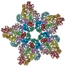
|
| 4 | x 6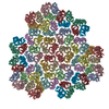
|
| 5 | 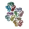
|
| 対称性 | 点対称性: (シェーンフリース記号: I (正20面体型対称)) |
-要素
| #1: タンパク質 | 分子量: 43346.219 Da / 分子数: 12 / 由来タイプ: 天然 / 由来: (天然) BACTERIOPHAGE PRD1 (ファージ) / 参照: UniProt: P22535 |
|---|
-実験情報
-実験
| 実験 | 手法: 電子顕微鏡法 |
|---|---|
| EM実験 | 試料の集合状態: PARTICLE / 3次元再構成法: 単粒子再構成法 |
- 試料調製
試料調製
| 構成要素 | 名称: BACTERIOPHAGE PRD1 / タイプ: VIRUS |
|---|---|
| 緩衝液 | 名称: TRISトリスヒドロキシメチルアミノメタン pH: 7.2 / 詳細: TRIS |
| 試料 | 濃度: 5 mg/ml / 包埋: NO / シャドウイング: NO / 染色: NO / 凍結: YES |
| 試料支持 | 詳細: HOLEY CARBON |
| 急速凍結 | 装置: HOMEMADE PLUNGER / 凍結剤: ETHANE / 詳細: ETHANE |
| 結晶化 | *PLUS 手法: cryo-electron microscopy |
- 電子顕微鏡撮影
| 顕微鏡 | モデル: FEI/PHILIPS CM200FEG / 日付: 2001年12月1日 |
|---|---|
| 電子銃 | 電子線源: FIELD EMISSION GUN / 加速電圧: 200 kV / 照射モード: FLOOD BEAM |
| 電子レンズ | モード: BRIGHT FIELDBright-field microscopy / 倍率(公称値): 50000 X / 倍率(補正後): 45300 X / 最大 デフォーカス(公称値): 4100 nm / 最小 デフォーカス(公称値): 1300 nm / Cs: 2 mm |
| 試料ホルダ | 温度: 95 K |
| 撮影 | 電子線照射量: 6 e/Å2 / フィルム・検出器のモデル: KODAK SO-163 FILM |
| 画像スキャン | デジタル画像の数: 13 |
| 放射波長 | 相対比: 1 |
- 解析
解析
| EMソフトウェア |
| ||||||||||||||||
|---|---|---|---|---|---|---|---|---|---|---|---|---|---|---|---|---|---|
| CTF補正 | 詳細: PHASE RESTORATION BY CTF- MULTIPLICATION OF IMAGES. AMPLITUDE RESTORATION BY COMPARISON WITH QUASI- ATOMIC MODEL | ||||||||||||||||
| 対称性 | 点対称性: I (正20面体型対称) | ||||||||||||||||
| 3次元再構成 | 手法: POLAR FOURIER TRANSFORM, CROSS- COMMON LINES / 解像度: 13.5 Å / 粒子像の数: 1468 / ピクセルサイズ(公称値): 3.68 Å / ピクセルサイズ(実測値): 3.34 Å / 倍率補正: COMPARISON WITH X- RAY DATA 詳細: RIGID BODY REFINEMENT AGAINST CRYO-EM MAP WAS DONE USING XPLOR 3.851 (BRUNGER) DATA USED IN REFINEMENT. NUMBER OF REFLECTIONS 1430672 対称性のタイプ: POINT | ||||||||||||||||
| 原子モデル構築 | プロトコル: RIGID BODY FIT / 空間: RECIPROCAL / Target criteria: R-factor / 詳細: METHOD--RIGID BODY REFINEMENT PROTOCOL--X-RAY | ||||||||||||||||
| 原子モデル構築 | PDB-ID: 1GW7 | ||||||||||||||||
| 精密化 | 最高解像度: 13.5 Å | ||||||||||||||||
| 精密化ステップ | サイクル: LAST / 最高解像度: 13.5 Å
|
