 Movie
Movie Controller

[English] 日本語
 Yorodumi
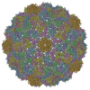
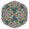
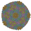
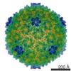
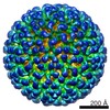
- PDB-1hb7: quasi-atomic resolution model of bacteriophage PRD1 sus1 mutant, ... -
+ Open data

- Basic information
| Entry | Database: PDB / ID: 1hb7 | ||||||
|---|---|---|---|---|---|---|---|
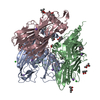
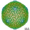
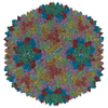
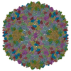
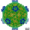
| Title | quasi-atomic resolution model of bacteriophage PRD1 sus1 mutant, obtained by combined cryo-EM and X-ray crystallography. | ||||||
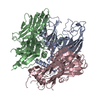
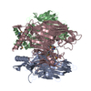
 Components Components | BACTERIOPHAGE PRD1 SUS1 MUTANT CAPSID | ||||||
 Keywords Keywords |  VIRUS / VIRUS/VIRAL PROTEIN / TECTIVIRIDAE / ICOSAHEDRAL VIRUS VIRUS / VIRUS/VIRAL PROTEIN / TECTIVIRIDAE / ICOSAHEDRAL VIRUS | ||||||
| Function / homology | Bacteriophage PRD1, P3 / Bacteriophage PRD1, P3, N-terminal / P3 major capsid protein / Group II dsDNA virus coat/capsid protein / Viral coat protein subunit / viral capsid / Major capsid protein P3 Function and homology information Function and homology information | ||||||
| Biological species |   BACTERIOPHAGE PRD1 (virus) BACTERIOPHAGE PRD1 (virus) | ||||||
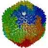
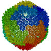
| Method | ELECTRON MICROSCOPY / single particle reconstruction / cryo EM / Resolution: 14 Å | ||||||
 Authors Authors | San Martin, C. / Burnett, R.M. / De Haas, F. / Heinkel, R. / Rutten, T. / Fuller, S.D. / Butcher, S.J. / Bamford, D.H. | ||||||
 Citation Citation | Journal: Structure / Year: 2001 Title: Combined EM/X-ray imaging yields a quasi-atomic model of the adenovirus-related bacteriophage PRD1 and shows key capsid and membrane interactions. Authors: C S Martín / R M Burnett / F de Haas / R Heinkel / T Rutten / S D Fuller / S J Butcher / D H Bamford /  Abstract: BACKGROUND: The dsDNA bacteriophage PRD1 has a membrane inside its icosahedral capsid. While its large size (66 MDa) hinders the study of the complete virion at atomic resolution, a 1.65-A ...BACKGROUND: The dsDNA bacteriophage PRD1 has a membrane inside its icosahedral capsid. While its large size (66 MDa) hinders the study of the complete virion at atomic resolution, a 1.65-A crystallographic structure of its major coat protein, P3, is available. Cryo-electron microscopy (cryo-EM) and three-dimensional reconstruction have shown the capsid at 20-28 A resolution. Striking architectural similarities between PRD1 and the mammalian adenovirus indicate a common ancestor. RESULTS: The P3 atomic structure has been fitted into improved cryo-EM reconstructions for three types of PRD1 particles: the wild-type virion, a packaging mutant without DNA, and a P3-shell lacking ...RESULTS: The P3 atomic structure has been fitted into improved cryo-EM reconstructions for three types of PRD1 particles: the wild-type virion, a packaging mutant without DNA, and a P3-shell lacking the membrane and the vertices. Establishing the absolute EM scale was crucial for an accurate match. The resulting "quasi-atomic" models of the capsid define the residues involved in the major P3 interactions, within the quasi-equivalent interfaces and with the membrane, and show how these are altered upon DNA packaging. CONCLUSIONS: The new cryo-EM reconstructions reveal the structure of the PRD1 vertex and the concentric packing of DNA. The capsid is essentially unchanged upon DNA packaging, with alterations ...CONCLUSIONS: The new cryo-EM reconstructions reveal the structure of the PRD1 vertex and the concentric packing of DNA. The capsid is essentially unchanged upon DNA packaging, with alterations limited to those P3 residues involved in membrane contacts. These are restricted to a few of the N termini along the icosahedral edges in the empty particle; DNA packaging leads to a 4-fold increase in the number of contacts, including almost all copies of the N terminus and the loop between the two beta barrels. Analysis of the P3 residues in each quasi-equivalent interface suggests two sites for minor proteins in the capsid edges, analogous to those in adenovirus. #1: Journal: Acta Crystallogr D Biol Crystallogr / Year: 2002Title: The X-ray crystal structure of P3, the major coat protein of the lipid-containing bacteriophage PRD1, at 1.65 A resolution. Authors: Stacy D Benson / Jaana K H Bamford / Dennis H Bamford / Roger M Burnett / Abstract: P3 has been imaged with X-ray crystallography to reveal a trimeric molecule with strikingly similar characteristics to hexon, the major coat protein of adenovirus. The structure of native P3 has now ...P3 has been imaged with X-ray crystallography to reveal a trimeric molecule with strikingly similar characteristics to hexon, the major coat protein of adenovirus. The structure of native P3 has now been extended to 1.65 A resolution (R(work) = 19.0% and R(free) = 20.8%). The new high-resolution model shows that P3 forms crystals through hydrophobic patches solvated by 2-methyl-2,4-pentanediol molecules. It reveals details of how the molecule's high stability may be achieved through ordered solvent in addition to intra- and intersubunit interactions. Of particular importance is a 'puddle' at the top of the molecule containing a four-layer deep hydration shell that cross-links a complex structural feature formed by 'trimerization loops'. These loops also link subunits by extending over a neighbor to reach the third subunit in the trimer. As each subunit has two eight-stranded viral jelly rolls, the trimer has a pseudo-hexagonal shape to allow close packing in its 240 hexavalent capsid positions. Flexible regions in P3 facilitate these interactions within the capsid and with the underlying membrane. A selenometh-ionine P3 derivative, with which the structure was solved, has been refined to 2.2 A resolution (R(work) = 20.1% and R(free) = 22.8%). The derivatized molecule is essentially unchanged, although synchrotron radiation has the curious effect of causing it to rotate about its threefold axis. P3 is a second example of a trimeric 'double-barrel' protein that forms a stable building block with optimal shape for constructing a large icosahedral viral capsid. A major difference is that hexon has long variable loops that distinguish different adenovirus species. The short loops in P3 and the severe constraints of its various interactions explain why the PRD1 family has highly conserved coat proteins. #2: Journal: Cell / Year: 1999Title: Viral evolution revealed by bacteriophage PRD1 and human adenovirus coat protein structures. Authors: S D Benson / J K Bamford / D H Bamford / R M Burnett / Abstract: The unusual bacteriophage PRD1 features a membrane beneath its icosahedral protein coat. The crystal structure of the major coat protein, P3, at 1.85 A resolution reveals a molecule with three ...The unusual bacteriophage PRD1 features a membrane beneath its icosahedral protein coat. The crystal structure of the major coat protein, P3, at 1.85 A resolution reveals a molecule with three interlocking subunits, each with two eight-stranded viral jelly rolls normal to the viral capsid, and putative membrane-interacting regions. Surprisingly, the P3 molecule closely resembles hexon, the equivalent protein in human adenovirus. Both viruses also have similar overall architecture, with identical capsid lattices and attachment proteins at their vertices. Although these two dsDNA viruses infect hosts from very different kingdoms, their striking similarities, from major coat protein through capsid architecture, strongly suggest their evolutionary relationship. #3: Journal: EMBO J / Year: 1995 Title: DNA packaging orders the membrane of bacteriophage PRD1. Authors: S J Butcher / D H Bamford / S D Fuller /  Abstract: Bacteriophage PRD1 contains a linear dsDNA genome enclosed by a lipid membrane lying within a protein coat. Determination of the structure of the detergent-treated particle to 2 nm by cryo-electron ...Bacteriophage PRD1 contains a linear dsDNA genome enclosed by a lipid membrane lying within a protein coat. Determination of the structure of the detergent-treated particle to 2 nm by cryo-electron microscopy and three-dimensional reconstruction has defined the position of the major coat protein P3. The coat contains 240 copies of trimeric P3 packed into positions of local 6-fold symmetry on a T = 25 lattice. The three-dimensional structures of the PRD1 virion and a DNA packaging mutant to a resolution of 2.8 nm have revealed specific interactions between the coat and the underlying membrane. The membrane is clearly visible as two leaflets separated by 2 nm and spanned by transmembrane density. The size of the coat does not change upon DNA packaging. Instead, the number of interactions seen between the protein shell and the membrane and the order of the membrane components increase. Thus the membrane of PRD1 plays a role in assembly which is akin to that played by the nucleocapsid in other membrane viruses. | ||||||
| History |
| ||||||
| Remark 700 | SHEET THE SHEET STRUCTURE OF THIS MOLECULE IS BIFURCATED. IN ORDER TO REPRESENT THIS FEATURE IN ... SHEET THE SHEET STRUCTURE OF THIS MOLECULE IS BIFURCATED. IN ORDER TO REPRESENT THIS FEATURE IN THE SHEET RECORDS BELOW, TWO SHEETS ARE DEFINED. |
- Structure visualization
| Movie |
Movie viewer |
|---|---|
| Structure viewer | Molecule: MolmilJmol/JSmol |
- Downloads & links
-Download
| PDBx/mmCIF format | 1hb7.cif.gz | 726.1 KB | Display | PDBx/mmCIF format |
|---|---|---|---|---|
| PDB format | pdb1hb7.ent.gz | 598.1 KB | Display | PDB format |
| PDBx/mmJSON format | 1hb7.json.gz | Tree view | PDBx/mmJSON format | |
| Others |  Other downloads Other downloads |
-Validation report
| Arichive directory | https://data.pdbj.org/pub/pdb/validation_reports/hb/1hb7ftp://data.pdbj.org/pub/pdb/validation_reports/hb/1hb7 | HTTPS FTP |
|---|
-Related structure data
| Related structure data | 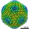 1013MC 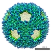 1014C 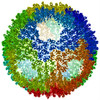 1hb5C 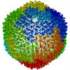 1hb9C C: citing same article ( M: map data used to model this data |
|---|---|
| Similar structure data |

-Links
 PDBj
PDBj

- Assembly
| Deposited unit | 
|
|---|---|
| 1 | x 60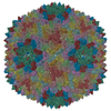
|
| 2 |
|
| 3 | x 5
|
| 4 | x 6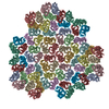
|
| 5 | 
|
| Symmetry | Point symmetry: (Schoenflies symbol: I (icosahedral)) |
-Components
| #1: Protein | Mass: 43346.219 Da / Num. of mol.: 12 Source method: isolated from a genetically manipulated source Source: (gene. exp.) BACTERIOPHAGE PRD1 (virus) / Strain: DS88 / Production host:  SALMONELLA TYPHIMURIUM (bacteria) / Strain (production host): DS88 / References: UniProt: P22535 SALMONELLA TYPHIMURIUM (bacteria) / Strain (production host): DS88 / References: UniProt: P22535Compound details | THE PRD1 SUS1 MUTANT LACKS THE PACKAGING PROTEIN P9 AND PRODUCES ONLY EMPTY PARTICLES, WHICH ...THE PRD1 SUS1 MUTANT LACKS THE PACKAGING PROTEIN P9 AND PRODUCES ONLY EMPTY PARTICLES, WHICH REPRESENT AN ASSEMBLY INTERMEDIA | |
|---|
-Experimental details
-Experiment
| Experiment | Method: ELECTRON MICROSCOPY |
|---|---|
| EM experiment | Aggregation state: PARTICLE / 3D reconstruction method: single particle reconstruction |
- Sample preparation
| Component | Name: BACTERIOPHAGE PRD1 SUS1 MUTANT / Type: VIRUS Details: 400 MESH COPPER GLOW DISCHARGE SAMPLES WERE PREPARED AS THIN LAYERS OF VITREOUS ICE |
|---|---|
| Buffer solution | pH: 7.2 |
| Specimen | Embedding applied: NO / Shadowing applied: NO / Staining applied: NO / Vitrification applied: YES |
| Specimen support | Details: HOLEY CARBON |
| Vitrification | Instrument: HOMEMADE PLUNGER / Cryogen name: ETHANE / Details: PLUNGE VITRIFICATION |
- Electron microscopy imaging
| Microscopy | Model: FEI/PHILIPS CM200FEG / Date: Jun 15, 1998 Details: SAMPLES WERE MAINTAINED AT LIQUID NITROGEN TEMPERATURES IN THE MICROSCOPE WITH A GATAN 626-0300 CRYOTRANSFER HOLDER |
|---|---|
| Electron gun | Electron source: FIELD EMISSION GUN / Accelerating voltage: 200 kV / Illumination mode: OTHER |
| Electron lens | Mode: BRIGHT FIELDBright-field microscopy / Nominal magnification: 36000 X / Nominal defocus max: 4100 nm / Nominal defocus min: 1300 nm / Cs: 2 mm |
| Specimen holder | Temperature: 95 K / Tilt angle max: 0 ° / Tilt angle min: 0 ° |
| Image recording | Electron dose: 10 e/Å2 / Film or detector model: KODAK SO-163 FILM |
| Image scans | Num. digital images: 29 |
| Radiation wavelength | Relative weight: 1 |
- Processing
| EM software |
| ||||||||||||
|---|---|---|---|---|---|---|---|---|---|---|---|---|---|
| Symmetry | Point symmetry: I (icosahedral) | ||||||||||||
| 3D reconstruction | Method: ICOSAHEDRALIcosahedron / Resolution: 14 Å / Num. of particles: 1800 / Nominal pixel size: 3.68 Å / Actual pixel size: 3.44 Å Magnification calibration: THE PIXEL SIZE OF THE CRYO-EM MAP WAS OBTAINED USING THE X-RAY STRUCTURE OF THE P3 TRIMER AS A REFERENCE. AFTER AN INITIAL FITTING USING THE NOMINAL PIXEL SIZE, THE P3 ...Magnification calibration: THE PIXEL SIZE OF THE CRYO-EM MAP WAS OBTAINED USING THE X-RAY STRUCTURE OF THE P3 TRIMER AS A REFERENCE. AFTER AN INITIAL FITTING USING THE NOMINAL PIXEL SIZE, THE P3 TRIMERS IN THE ICOSAHEDRAL ASYMMETRIC UNIT WERE GRADUALLY TRANSLATED TOWARDS THE CENTER OF THE PARTICLE UNTIL THE CRYSTALLOGRAPHIC R-FACTOR WAS MINIMISED. Details: THE ORIENTATIONS WERE REFINED BY THE CROSS COMMON LINES LINES METHOD (SIMPLEX) AND THE POLAR FOURIER TRANSFORM METHOD. MODEL-BASED, POLAR-FOURIER-TRANSFORM (FULLER ET AL. 1996, J.STRUC.BIOL. ...Details: THE ORIENTATIONS WERE REFINED BY THE CROSS COMMON LINES LINES METHOD (SIMPLEX) AND THE POLAR FOURIER TRANSFORM METHOD. MODEL-BASED, POLAR-FOURIER-TRANSFORM (FULLER ET AL. 1996, J.STRUC.BIOL. 116, 48-55; BAKER AND CHENG, 1996, J.STRUC.BIOL. 116, 120-130) MODEL-BASED CROSS COMMON LINES SEARCH AND REFINEMENT (CROWTHER ET AL. 1970, NATURE (LONDON) 226, 421-425; FULLER ET AL. 1996, J.STRUC.BIOL. 116, 48-55; FERLENGHI ET AL. 1998, J.MOL.BIOL. 283, 71-81). THE EFFECTIVE RESOLUTION OF THE FINAL RECONSTRUCTED DENSITY WAS DETERMINED TO BE AT LEAST 25 ANGSTROMS, AS MEASURED BY RANDOMLY SPLITTING THE PARTICLES INTO TWO SETS AND CALCULATING THE FOURIER SHELL CORRELATION OBTAINED FROM SEPARATE RECONSTRUCTIONS (HARAUZ AND VAN HEEL 1986, OPTIK 73, 146-156). THE EIGENVALUE SPECTRUM GAVE AN INDICATION OF THE RANDOMNESS OF THE DATA THAT WAS INCLUDED IN THE RECONSTRUCTION. THE COMPLETENESS OF THE DATA WAS VERIFIED IN THAT ALL EIGENVALUES EXCEEDED 100. THE COORDINATES ARE IN THE P, Q, R FRAME IN ANGSTROM UNITS AND CORRESPOND TO ICOSAHEDRAL SYMMETRY AXES. THE ORIGIN IS CHOSEN AT THE CENTER OF THE VIRUS WITH P, Q AND R ALONG MUTUALLY PERPENDICULAR TWO-FOLD AXES OF THE ICOSAHEDRON. THEY SHOULD REMAIN IN THAT FRAME FOR THE EASE OF THE USER IN CREATING THE BIOLOGICALLY SIGNIFICANT VIRAL COMPLEX PARTICLE USING THE 60 ICOSAHEDRAL SYMMETRY OPERATORS. RESIDUES NOT VISIBLE IN THE ORIGINAL CRYSTAL STRUCTURES ARE NOT INCLUDED IN THE CRYO-EM STRUCTURE MODEL. Symmetry type: POINT | ||||||||||||
| Atomic model building | Protocol: RIGID BODY FIT / Space: RECIPROCAL / Target criteria: R-factor Details: METHOD--THE CRYSTAL STRUCTURE OF THE MAJOR COAT PROTEIN P3 (PDB FILE 1HX6) WAS PLACED INTO THE CRYO-EM DENSITY MAP. THE CAPSID PROTEIN WAS FIRST MANUALLY POSITIONED INTO THE CRYO-EM DENSITY ...Details: METHOD--THE CRYSTAL STRUCTURE OF THE MAJOR COAT PROTEIN P3 (PDB FILE 1HX6) WAS PLACED INTO THE CRYO-EM DENSITY MAP. THE CAPSID PROTEIN WAS FIRST MANUALLY POSITIONED INTO THE CRYO-EM DENSITY CORRESPONDING TO POSITIONS OF THE FOUR INDEPENDENT TRIMERS IN THE ICOSAHEDRAL ASYMMETRIC UNIT. THESE POSITIONS WERE THEN REFINED BY RIGID BODY REFINEMENT IN RECIPROCAL SPACE WITH THE PROGRAM XPLOR. QUALITY OF THE FIT R- FACTOR= 0.339, CROSS-CORRELATI0N COEFFICIENT 0.915, ATOMS OUTSIDE DENSITY PER ICOSAHEDRAL ASYMMETRIC UNIT 527 (1.5%), ATOM CLASHES PER ICOSAHEDRAL ASYMMETRIC UNIT 115 (0.3%) REFINEMENT PROTOCOL--RIGID BODY REFINEMENT | ||||||||||||
| Atomic model building | PDB-ID: 1HX6 | ||||||||||||
| Refinement | Highest resolution: 14 Å | ||||||||||||
| Refinement step | Cycle: LAST / Highest resolution: 14 Å
|
