 Movie
Movie Controller

[English] 日本語
 Yorodumi
- EMDB-1011: Minor proteins, mobile arms and membrane-capsid interactions in t... -
+ Open data

- Basic information
| Entry | Database: EMDB / ID: EMD-1011 | |||||||||
|---|---|---|---|---|---|---|---|---|---|---|
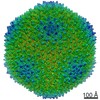
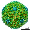
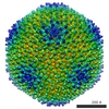
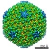
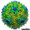
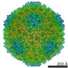
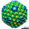
| Title | Minor proteins, mobile arms and membrane-capsid interactions in the bacteriophage PRD1 capsid. | |||||||||
 Map data Map data | This map is one of four described in San Martin, C., Huiskonen, J. T., Bamford, J. K., Butcher, S. J., Fuller, S. D., Bamford, D. H., and Burnett, R. M. (2002). Minor proteins, mobile arms and membrane-capsid interactions in the bacteriophage PRD1 capsid. Nat Struct Biol 9, 756-763. The other three maps are also available. | |||||||||
 Sample Sample |
| |||||||||
| Function / homology | Bacteriophage PRD1, P3 / Bacteriophage PRD1, P3, N-terminal / P3 major capsid protein / Group II dsDNA virus coat/capsid protein / Viral coat protein subunit / viral capsid / Major capsid protein P3 Function and homology information Function and homology information | |||||||||
| Biological species |   Enterobacteria phage PRD1 (virus) Enterobacteria phage PRD1 (virus) | |||||||||
| Method | single particle reconstruction / cryo EM / Resolution: 16.0 Å | |||||||||
 Authors Authors | San Martin C / Huiskonen JT / Bamford JK / Butcher SJ / Fuller SD / Bamford DH / Burnett RM | |||||||||
 Citation Citation | Journal: Nat Struct Biol / Year: 2002 Title: Minor proteins, mobile arms and membrane-capsid interactions in the bacteriophage PRD1 capsid. Authors: Carmen San Martín / Juha T Huiskonen / Jaana K H Bamford / Sarah J Butcher / Stephen D Fuller / Dennis H Bamford / Roger M Burnett /  Abstract: Bacteriophage PRD1 shares many structural and functional similarities with adenovirus. A major difference is the PRD1 internal membrane, which acts in concert with vertex proteins to translocate the ...Bacteriophage PRD1 shares many structural and functional similarities with adenovirus. A major difference is the PRD1 internal membrane, which acts in concert with vertex proteins to translocate the phage genome into the host. Multiresolution models of the PRD1 capsid, together with genetic analyses, provide fine details of the molecular interactions associated with particle stability and membrane dynamics. The N- and C-termini of the major coat protein (P3), which are required for capsid assembly, act as conformational switches bridging capsid to membrane and linking P3 trimers. Electrostatic P3-membrane interactions increase virion stability upon DNA packaging. Newly revealed proteins suggest how the metastable vertex works and how the capsid edges are stabilized. | |||||||||
| History |
|
- Structure visualization
| Movie |
Movie viewer |
|---|---|
| Structure viewer | EM map: SurfViewMolmilJmol/JSmol |
| Supplemental images |

- Downloads & links
-EMDB archive
| Map data | emd_1011.map.gz | 59.1 MB | EMDB map data format | |
|---|---|---|---|---|
| Header (meta data) | emd-1011-v30.xmlemd-1011.xml | 19.9 KB 19.9 KB | Display Display | EMDB header |
| Images |  1011.gif 1011.gif | 24.9 KB | ||
| Others | emd_1011_additional_1.map.gzemd_1011_additional_2.map.gzemd_1011_additional_3.map.gz | 237.6 KB 237.8 KB 237.6 KB | ||
| Archive directory |  http://ftp.pdbj.org/pub/emdb/structures/EMD-1011ftp://ftp.pdbj.org/pub/emdb/structures/EMD-1011 http://ftp.pdbj.org/pub/emdb/structures/EMD-1011ftp://ftp.pdbj.org/pub/emdb/structures/EMD-1011 | HTTPS FTP |
-Validation report
| Summary document | emd_1011_validation.pdf.gz | 306.5 KB | Display | EMDB validaton report |
|---|---|---|---|---|
| Full document | emd_1011_full_validation.pdf.gz | 305.6 KB | Display | |
| Data in XML | emd_1011_validation.xml.gz | 6.7 KB | Display | |
| Arichive directory | https://ftp.pdbj.org/pub/emdb/validation_reports/EMD-1011ftp://ftp.pdbj.org/pub/emdb/validation_reports/EMD-1011 | HTTPS FTP |
-Related structure data
| Related structure data | 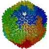 1gw7MC  1012C 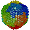 1gw8C M: atomic model generated by this map C: citing same article ( |
|---|---|
| Similar structure data |
-Links
| EMDB pages | EMDB (EBI/PDBe) / EMDataResource |
|---|---|
| Related items in Molecule of the Month |


-Map
| File | Download / File: emd_1011.map.gz / Format: CCP4 / Size: 62.5 MB / Type: IMAGE STORED AS FLOATING POINT NUMBER (4 BYTES) | ||||||||||||||||||||||||||||||||||||||||||||||||||||||||||||
|---|---|---|---|---|---|---|---|---|---|---|---|---|---|---|---|---|---|---|---|---|---|---|---|---|---|---|---|---|---|---|---|---|---|---|---|---|---|---|---|---|---|---|---|---|---|---|---|---|---|---|---|---|---|---|---|---|---|---|---|---|---|
| Annotation | This map is one of four described in San Martin, C., Huiskonen, J. T., Bamford, J. K., Butcher, S. J., Fuller, S. D., Bamford, D. H., and Burnett, R. M. (2002). Minor proteins, mobile arms and membrane-capsid interactions in the bacteriophage PRD1 capsid. Nat Struct Biol 9, 756-763. The other three maps are also available. | ||||||||||||||||||||||||||||||||||||||||||||||||||||||||||||
| Voxel size | X=Y=Z: 3.44 Å | ||||||||||||||||||||||||||||||||||||||||||||||||||||||||||||
| Density |
| ||||||||||||||||||||||||||||||||||||||||||||||||||||||||||||
| Symmetry | Space group: 1 | ||||||||||||||||||||||||||||||||||||||||||||||||||||||||||||
| Details | EMDB XML:
CCP4 map header:
| ||||||||||||||||||||||||||||||||||||||||||||||||||||||||||||
-Supplemental data
-Supplemental map: emd 1011 additional 1.map
| File | emd_1011_additional_1.map | ||||||||||||
|---|---|---|---|---|---|---|---|---|---|---|---|---|---|
| Projections & Slices |
| ||||||||||||
| Density Histograms |
 Z
Z Y
Y X
X


-Supplemental map: emd 1011 additional 2.map
| File | emd_1011_additional_2.map | ||||||||||||
|---|---|---|---|---|---|---|---|---|---|---|---|---|---|
| Projections & Slices |
| ||||||||||||
| Density Histograms |


-Supplemental map: emd 1011 additional 3.map
| File | emd_1011_additional_3.map | ||||||||||||
|---|---|---|---|---|---|---|---|---|---|---|---|---|---|
| Projections & Slices |
| ||||||||||||
| Density Histograms |


- Sample components
-Entire : Bacteriophage PRD1
| Entire | Name: Bacteriophage PRD1 (virus) |
|---|---|
| Components |
|
-Supramolecule #1000: Bacteriophage PRD1
| Supramolecule | Name: Bacteriophage PRD1 / type: sample / ID: 1000 Details: The sample is a virion containing at least 19 structural proteins and a double stranded DNA genome Oligomeric state: A pseudo T=25 assembly / Number unique components: 1 |
|---|---|
| Molecular weight | Theoretical: 70 MDa |
-Supramolecule #1: Enterobacteria phage PRD1
| Supramolecule | Name: Enterobacteria phage PRD1 / type: virus / ID: 1 / Name.synonym: bacteriophage PRD1 / Details: a T=25 virion / NCBI-ID: 10658 / Sci species name: Enterobacteria phage PRD1 / Virus type: VIRION / Virus isolate: STRAIN / Virus enveloped: No / Virus empty: No / Syn species name: bacteriophage PRD1 |
|---|---|
| Host (natural) | Organism:  Salmonella enterica (bacteria) / synonym: BACTERIA(EUBACTERIA) Salmonella enterica (bacteria) / synonym: BACTERIA(EUBACTERIA) |
| Virus shell | Shell ID: 1 / Name: P3 / Diameter: 70 Å / T number (triangulation number): 25 |
-Experimental details
-Structure determination
| Method | cryo EM |
|---|---|
 Processing Processing | single particle reconstruction |
| Aggregation state | particle |
-Sample preparation
| Concentration | 1 mg/mL |
|---|---|
| Buffer | pH: 7.2 / Details: 20 mM Tris HCl |
| Vitrification | Cryogen name: ETHANE / Chamber humidity: 60 % / Chamber temperature: 23 K / Instrument: HOMEMADE PLUNGER Details: Vitrification instrument: EMBL plunger. vitrification carried out at 23 degrees at ambient humidity Method: Blot for 1 s before plunging into ethane slush |
- Electron microscopy
| Microscope | FEI/PHILIPS CM200FEG/ST |
|---|---|
| Temperature | Average: 105 K |
| Image recording | Category: FILM / Film or detector model: KODAK SO-163 FILM / Digitization - Scanner: ZEISS SCAI / Digitization - Sampling interval: 7.0 µm / Number real images: 50 / Average electron dose: 6 e/Å2 / Camera length: 44 Details: images were scanned at 7 micron steps size and then averaged to give a final size of 14 microns Od range: 1 / Bits/pixel: 8 |
| Electron beam | Acceleration voltage: 200 kV / Electron source:  FIELD EMISSION GUN FIELD EMISSION GUN |
| Electron optics | Illumination mode: FLOOD BEAM / Imaging mode: BRIGHT FIELD / Cs: 2 mm / Nominal defocus max: 4.1 µm / Nominal defocus min: 1.3 µm / Nominal magnification: 36000 |
| Sample stage | Specimen holder: eucentric / Specimen holder model: GATAN LIQUID NITROGEN |
-Image processing
| Details | The particles were purified by rate-zonal centrifugation and ion-exchange chromatography Walin,Tuma,Thomas and Bamford Virology (1994) 201:1-7 |
|---|---|
| CTF correction | Details: normalized sum of ctf multiplied images |
| Final reconstruction | Applied symmetry - Point group: I (icosahedral) / Algorithm: OTHER / Resolution.type: BY AUTHOR / Resolution: 16.0 Å / Resolution method: OTHER / Software - Name: EMBL-ICOS, MRC Details: final maps were calculated by making a normalized sum of seperate ctf multiplied maps Baker, T. S., Olson, N. H., and Fuller, S. D. (1999). Adding the third dimension to virus life cycles: ...Details: final maps were calculated by making a normalized sum of seperate ctf multiplied maps Baker, T. S., Olson, N. H., and Fuller, S. D. (1999). Adding the third dimension to virus life cycles: Three-Dimensional Reconstruction of Icosahedral Viruses from Cryo-Electron Micrographs. Microbiology and Molecular Biology Reviews 63, 862-922. Butcher, S. J., Bamford, D. H., and Fuller, S. D. (1995). DNA packaging orders the membrane of bacteriophage PRD1. Embo J 14, 6078-6086. Ferlenghi, I., Gowen, B., de Haas, F., Mancini, E. J., Garoff, H., Sjoberg, M., and Fuller, S. D. (1998). The first step: activation of the Semliki Forest virus spike protein precursor causes a localized conformational change in the trimeric spike. J Mol Biol 283, 71-81. Fuller, S. D., Berriman, J. A., Butcher, S. J., and Gowen, B. E. (1995). Low pH induces swiveling of the glycoprotein heterodimers in the Semliki Forest virus spike complex. Cell 81, 715-725. Fuller, S. D., Butcher, S. J., Cheng, R. H., and Baker, T. S. (1996). Three-dimensional reconstruction of icosahedral particles--the uncommon line. J Struct Biol 116, 48-55. Mancini, E. J., Clarke, M., Gowen, B., Rutten, T., and Fuller, S. D. (2000). Cryo-electron microscopy reveals the functional organization of an enveloped virus, Semliki Forest virus. Molecular Cell 5, 255-266. Mancini, E. J., de Haas, F., and Fuller, S. D. (1997). High-resolution icosahedral reconstruction: fulfilling the promise of cryo-electron microscopy. Structure 5, 741-750. San Martin, C., Burnett, R. M., de Haas, F., Heinkel, R., Rutten, T., Fuller, S. D., Butcher, S. J., and Bamford, D. H. (2001). Combined EM/X-ray imaging yields a quasi-atomic model of the adenovirus-related bacteriophage PRD1 and shows key capsid and membrane interactions. Structure (Camb) 9, 917-930. San Martin, C., Huiskonen, J. T., Bamford, J. K., Butcher, S. J., Fuller, S. D., Bamford, D. H., and Burnett, R. M. (2002). Minor proteins, mobile arms and membrane-capsid interactions in the bacteriophage PRD1 capsid. Nat Struct Biol 9, 756-763. Sheehan, B., Fuller, S. D., Pique, M. E., and Yeager, M. (1996). AVS software for visualization in molecular microscopy. J Struct Biol 116, 99-106. Number images used: 1468 |
| Final angle assignment | Details: range giving min eigenvalues for inversion of less than .01 |
-Atomic model buiding 1
| Initial model | PDB ID: Chain - #0 - Chain ID: A / Chain - #1 - Chain ID: B / Chain - #2 - Chain ID: C / Chain - #3 - Chain ID: D / Chain - #4 - Chain ID: E / Chain - #5 - Chain ID: F / Chain - #6 - Chain ID: G / Chain - #7 - Chain ID: H / Chain - #8 - Chain ID: I |
|---|---|
| Software | Name: emfit (M. Rossmann Cheng, R., Kuhn, R., Olson, N., Rossmann, M., and Baker, T. (1995). Nucleocapsid and glycoprotein organization in an enveloped virus. Cell 80, 621-630.) |
| Details | Protocol: real space refinement. The scale of the map and the effective resolution were determined from a comparison between the P3 portion of the WT reconstruction and the atomic model |
| Refinement | Space: REAL / Protocol: RIGID BODY FIT / Target criteria: minimizing R factor (final 47.7%) |
| Output model | PDB-1gw7: |
