misfolded RNA binding / Group I intron splicing / RNA folding / positive regulation of ribosome biogenesis / DnaA-L2 complex / negative regulation of translational initiation / negative regulation of DNA-templated DNA replication initiation / mRNA regulatory element binding translation repressor activity / assembly of large subunit precursor of preribosome / positive regulation of RNA splicing ...misfolded RNA binding / Group I intron splicing / RNA folding / positive regulation of ribosome biogenesis / DnaA-L2 complex / negative regulation of translational initiation / negative regulation of DNA-templated DNA replication initiation / mRNA regulatory element binding translation repressor activity / assembly of large subunit precursor of preribosome / positive regulation of RNA splicing / cytosolic ribosome assembly / transcription antitermination / regulation of cell growth / maintenance of translational fidelity / DNA-templated transcription termination / mRNA 5'-UTR binding / ribosomal small subunit biogenesis / ribosomal large subunit assembly / small ribosomal subunit rRNA binding / ribosomal small subunit assembly / cytosolic small ribosomal subunit / ribosome binding / large ribosomal subunit / small ribosomal subunit / cytoplasmic translation / 5S rRNA binding / cytosolic large ribosomal subunit / transferase activity / tRNA binding / negative regulation of translation / rRNA binding / リボソーム / structural constituent of ribosome / 翻訳 (生物学) / ribonucleoprotein complex / response to antibiotic / mRNA binding / RNA binding / zinc ion binding / 生体膜 / metal ion binding / 細胞質基質 / 細胞質 類似検索 - 分子機能
Ribosomal protein S21, conserved site / Ribosomal protein S21 signature. / Ribosomal protein L25, short-form / Ribosomal protein S14, bacterial/plastid / Ribosomal protein L31 type A / Ribosomal protein S21 superfamily / Ribosomal protein S21 / Ribosomal protein S16, conserved site / Ribosomal protein S16 signature. / Ribosomal protein S21 ...Ribosomal protein S21, conserved site / Ribosomal protein S21 signature. / Ribosomal protein L25, short-form / Ribosomal protein S14, bacterial/plastid / Ribosomal protein L31 type A / Ribosomal protein S21 superfamily / Ribosomal protein S21 / Ribosomal protein S16, conserved site / Ribosomal protein S16 signature. / Ribosomal protein S21 / Ribosomal protein L31 signature. / Ribosomal protein L31 / Ribosomal protein L31 superfamily / Ribosomal protein L31 / Ribosomal protein L21, conserved site / Ribosomal protein L21 signature. / Ribosomal protein L16 signature 1. / : / Ribosomal protein L6, conserved site / Ribosomal protein L6 signature 1. / Ribosomal protein L16, conserved site / Ribosomal protein L16 signature 2. / Ribosomal protein L9 signature. / Ribosomal protein L17 signature. / Ribosomal protein L9, bacteria/chloroplast / Ribosomal protein L9, C-terminal / Ribosomal protein L9, C-terminal domain / Ribosomal protein L9, C-terminal domain superfamily / Ribosomal L25p family / Ribosomal protein L25 / Ribosomal protein L28/L24 superfamily / Ribosomal protein L36 signature. / Ribosomal protein L25/Gln-tRNA synthetase, N-terminal / Ribosomal protein L32p, bacterial type / Ribosomal protein L25/Gln-tRNA synthetase, anti-codon-binding domain superfamily / Ribosomal protein L9, N-terminal domain superfamily / Ribosomal protein L9 / Ribosomal protein L9, N-terminal / Ribosomal protein L9, N-terminal domain / Ribosomal protein L28 / Ribosomal protein L35, conserved site / Ribosomal protein L35 signature. / Ribosomal protein L33, conserved site / Ribosomal protein L33 signature. / Ribosomal protein L35, non-mitochondrial / Ribosomal protein L5, bacterial-type / Ribosomal protein L6, bacterial-type / Ribosomal protein L18, bacterial-type / Ribosomal protein L19, conserved site / Ribosomal protein L19 signature. / Ribosomal protein L36 / Ribosomal protein L36 superfamily / Ribosomal protein L36 / Ribosomal protein L9/RNase H1, N-terminal / Ribosomal protein L20 signature. / Ribosomal protein S3, bacterial-type / Ribosomal protein S6, conserved site / Ribosomal protein S6 signature. / Ribosomal protein L27, conserved site / Ribosomal protein L27 signature. / Ribosomal protein S19, bacterial-type / Ribosomal protein S7, bacterial/organellar-type / Ribosomal protein S11, bacterial-type / Ribosomal protein S13, bacterial-type / Ribosomal protein S20 / Ribosomal protein S20 superfamily / Ribosomal protein S20 / Ribosomal protein S9, bacterial/plastid / Ribosomal protein L14P, bacterial-type / Ribosomal protein S4, bacterial-type / Ribosomal protein L34, conserved site / Ribosomal protein L34 signature. / 30S ribosomal protein S17 / Ribosomal protein S5, bacterial-type / Ribosomal protein S6, plastid/chloroplast / Ribosomal protein L35 / Ribosomal protein L35 superfamily / Ribosomal protein L2, bacterial/organellar-type / Ribosomal protein L35 / Ribosomal protein S2, bacteria/mitochondria/plastid / Ribosomal L28 family / Ribosomal protein L33 / Ribosomal protein L33 / Ribosomal protein L28/L24 / Ribosomal protein L33 superfamily / Ribosomal protein L30, bacterial-type / Ribosomal protein L16 / Ribosomal protein L18 / Ribosomal L18 of archaea, bacteria, mitoch. and chloroplast / Ribosomal protein S18, conserved site / Ribosomal protein S18 signature. / L28p-like / Ribosomal protein L20 / Ribosomal protein S16 / Ribosomal protein S16 / Ribosomal protein S16 domain superfamily / Ribosomal protein L20 / Ribosomal protein L20, C-terminal / Ribosomal protein L21 / Ribosomal protein L27 類似検索 - ドメイン・相同性
Large ribosomal subunit protein uL15 / Small ribosomal subunit protein bS18 / Large ribosomal subunit protein bL36 / Small ribosomal subunit protein bS21 / Large ribosomal subunit protein bL28 / Large ribosomal subunit protein uL24 / Large ribosomal subunit protein bL17 / 30S ribosomal protein S9 / Small ribosomal subunit protein uS13 / 30S ribosomal protein S17 ...Large ribosomal subunit protein uL15 / Small ribosomal subunit protein bS18 / Large ribosomal subunit protein bL36 / Small ribosomal subunit protein bS21 / Large ribosomal subunit protein bL28 / Large ribosomal subunit protein uL24 / Large ribosomal subunit protein bL17 / 30S ribosomal protein S9 / Small ribosomal subunit protein uS13 / 30S ribosomal protein S17 / Large ribosomal subunit protein uL5 / Large ribosomal subunit protein uL23 / Large ribosomal subunit protein bL21 / 50S ribosomal protein L22 / 50S ribosomal protein L6 / Large ribosomal subunit protein bL20 / Small ribosomal subunit protein uS11 / Small ribosomal subunit protein uS3 / Large ribosomal subunit protein bL34 / Small ribosomal subunit protein bS16 / Small ribosomal subunit protein bS20 / Large ribosomal subunit protein uL29 / Large ribosomal subunit protein bL25 / Large ribosomal subunit protein bL32 / Small ribosomal subunit protein uS8 / 30S ribosomal protein S2 / Small ribosomal subunit protein uS15 / Large ribosomal subunit protein bL33 / 50S ribosomal protein L16 / 50S ribosomal protein L31 / 50S ribosomal protein L18 / 50S ribosomal protein L4 / Small ribosomal subunit protein bS6 / Small ribosomal subunit protein uS7 / Large ribosomal subunit protein bL27 / Small ribosomal subunit protein uS12 / Small ribosomal subunit protein uS4 / Small ribosomal subunit protein uS5 / Large ribosomal subunit protein uL2 / Large ribosomal subunit protein uL3 / Small ribosomal subunit protein uS19 / Large ribosomal subunit protein uL13 / 50S ribosomal protein L35 / Large ribosomal subunit protein uL14 / Small ribosomal subunit protein uS14 / Large ribosomal subunit protein bL19 / Small ribosomal subunit protein uS10 / Large ribosomal subunit protein uL30 / Large ribosomal subunit protein bL9 類似検索 - 構成要素
National Institutes of Health/National Institute Of Allergy and Infectious Diseases (NIH/NIAID)
R01AI137270
National Institutes of Health/National Institute Of Allergy and Infectious Diseases (NIH/NIAID)
F32AI148120
Other private
Sanghvi-Agarwal Innovation Award
Other government
NSF GRFP (1650113)
Other government
UCSF Discovery Fellowship
引用
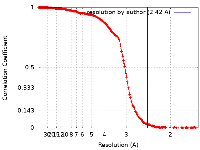
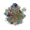
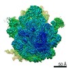
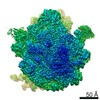
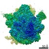
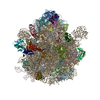
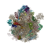
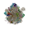
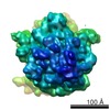
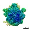
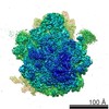
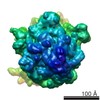
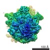
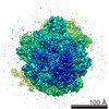
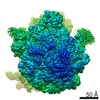
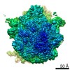
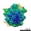
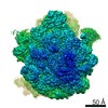
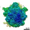
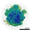
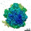
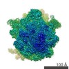
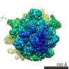
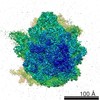
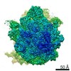
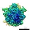
ジャーナル: Nat Struct Mol Biol / 年: 2022 タイトル: Structural basis for context-specific inhibition of translation by oxazolidinone antibiotics. 著者: Kaitlyn Tsai / Vanja Stojković / D John Lee / Iris D Young / Teresa Szal / Dorota Klepacki / Nora Vázquez-Laslop / Alexander S Mankin / James S Fraser / Danica Galonić Fujimori / 要旨: The antibiotic linezolid, the first clinically approved member of the oxazolidinone class, inhibits translation of bacterial ribosomes by binding to the peptidyl transferase center. Recent work has ...The antibiotic linezolid, the first clinically approved member of the oxazolidinone class, inhibits translation of bacterial ribosomes by binding to the peptidyl transferase center. Recent work has demonstrated that linezolid does not inhibit peptide bond formation at all sequences but rather acts in a context-specific manner, namely when alanine occupies the penultimate position of the nascent chain. However, the molecular basis for context-specificity has not been elucidated. Here we show that the second-generation oxazolidinone radezolid also induces stalling with a penultimate alanine, and we determine high-resolution cryo-EM structures of linezolid- and radezolid-stalled ribosome complexes to explain their mechanism of action. These structures reveal that the alanine side chain fits within a small hydrophobic crevice created by oxazolidinone, resulting in improved ribosome binding. Modification of the ribosome by the antibiotic resistance enzyme Cfr disrupts stalling due to repositioning of the modified nucleotide. Together, our findings provide molecular understanding for the context-specificity of oxazolidinones.
 ムービー
ムービー コントローラー


 データを開く
 基本情報
基本情報 マップデータ
 試料
試料 キーワード
キーワード oxazolidinone /
  機能・相同性情報
 データ登録者
 引用
引用
 構造の表示
構造の表示
 ダウンロードとリンク
 emd_24804.png
 http://ftp.pdbj.org/pub/emdb/structures/EMD-24804


 試料の構成要素

 解析
解析 電子顕微鏡法