ムービー
ムービー コントローラー


+ データを開く

- 基本情報
基本情報
| 登録情報 | データベース: EMDB / ID: EMD-2392 | |||||||||
|---|---|---|---|---|---|---|---|---|---|---|
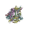
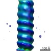
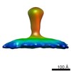
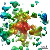
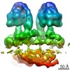
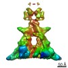
| タイトル | Electron cryomicroscopy of human Respiratory Syncytial Virus prefusion F | |||||||||
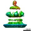
 マップデータ マップデータ | Subtomogram average of hRSV prefusion F | |||||||||
 試料 試料 |
| |||||||||
 キーワード キーワード | RSV /  fusion protein (融合タンパク質) / prefusion fusion protein (融合タンパク質) / prefusion | |||||||||
| 機能・相同性 |  機能・相同性情報 機能・相同性情報positive regulation of syncytium formation by virus / host cell Golgi membrane / entry receptor-mediated virion attachment to host cell / symbiont entry into host cell / fusion of virus membrane with host plasma membrane / エンベロープ (ウイルス) / host cell plasma membrane / virion membrane / 生体膜 / identical protein binding類似検索 - 分子機能 | |||||||||
| 生物種 |  Human respiratory syncytial virus (RSウイルス) Human respiratory syncytial virus (RSウイルス) | |||||||||
| 手法 | サブトモグラム平均法 / クライオ電子顕微鏡法 / 解像度: 47.0 Å | |||||||||
 データ登録者 データ登録者 | Liljeroos L / Krzyzaniak MA / Helenius A / Butcher SJ | |||||||||
 引用 引用 | ジャーナル: Proc Natl Acad Sci U S A / 年: 2013 タイトル: Architecture of respiratory syncytial virus revealed by electron cryotomography. 著者: Lassi Liljeroos / Magdalena Anna Krzyzaniak / Ari Helenius / Sarah Jane Butcher /  要旨: Human respiratory syncytial virus is a human pathogen that causes severe infection of the respiratory tract. Current information about the structure of the virus and its interaction with host cells ...Human respiratory syncytial virus is a human pathogen that causes severe infection of the respiratory tract. Current information about the structure of the virus and its interaction with host cells is limited. We carried out an electron cryotomographic characterization of cell culture-grown human respiratory syncytial virus to determine the architecture of the virion. The particles ranged from 100 nm to 1,000 nm in diameter and were spherical, filamentous, or a combination of the two. The filamentous morphology correlated with the presence of a cylindrical matrix protein layer linked to the inner leaflet of the viral envelope and with local ordering of the glycoprotein spikes. Recombinant viruses with only the fusion protein in their envelope showed that these glycoproteins were predominantly in the postfusion conformation, but some were also in the prefusion form. The ribonucleocapsids were left-handed, randomly oriented, and curved inside the virions. In filamentous particles, they were often adjacent to an intermediate layer of protein assigned to M2-1 (an envelope-associated protein known to mediate association of ribonucleocapsids with the matrix protein). Our results indicate important differences in structure between the Paramyxovirinae and Pneumovirinae subfamilies within the Paramyxoviridae, and provide fresh insights into host cell exit of a serious pathogen. | |||||||||
| 履歴 |
|
- 構造の表示
構造の表示
| ムービー |
ムービービューア |
|---|---|
| 構造ビューア | EMマップ: SurfViewMolmilJmol/JSmol |
| 添付画像 |
- ダウンロードとリンク
-EMDBアーカイブ
| マップデータ | emd_2392.map.gz | 952.8 KB | EMDBマップデータ形式 | |
|---|---|---|---|---|
| ヘッダ (付随情報) | emd-2392-v30.xmlemd-2392.xml | 8.9 KB 8.9 KB | 表示 表示 | EMDBヘッダ |
| 画像 | emd_2392.tif | 31.8 KB | ||
| アーカイブディレクトリ |  http://ftp.pdbj.org/pub/emdb/structures/EMD-2392ftp://ftp.pdbj.org/pub/emdb/structures/EMD-2392 http://ftp.pdbj.org/pub/emdb/structures/EMD-2392ftp://ftp.pdbj.org/pub/emdb/structures/EMD-2392 | HTTPS FTP |
-関連構造データ
-リンク
| EMDBのページ | EMDB (EBI/PDBe) / EMDataResource |
|---|---|
| 「今月の分子」の関連する項目 |


-マップ
| ファイル | ダウンロード / ファイル: emd_2392.map.gz / 形式: CCP4 / 大きさ: 1001 KB / タイプ: IMAGE STORED AS FLOATING POINT NUMBER (4 BYTES) | ||||||||||||||||||||||||||||||||||||||||||||||||||||||||||||||||||||
|---|---|---|---|---|---|---|---|---|---|---|---|---|---|---|---|---|---|---|---|---|---|---|---|---|---|---|---|---|---|---|---|---|---|---|---|---|---|---|---|---|---|---|---|---|---|---|---|---|---|---|---|---|---|---|---|---|---|---|---|---|---|---|---|---|---|---|---|---|---|
| 注釈 | Subtomogram average of hRSV prefusion F | ||||||||||||||||||||||||||||||||||||||||||||||||||||||||||||||||||||
| ボクセルのサイズ | X=Y=Z: 7.68 Å | ||||||||||||||||||||||||||||||||||||||||||||||||||||||||||||||||||||
| 密度 |
| ||||||||||||||||||||||||||||||||||||||||||||||||||||||||||||||||||||
| 対称性 | 空間群: 1 | ||||||||||||||||||||||||||||||||||||||||||||||||||||||||||||||||||||
| 詳細 | EMDB XML:
CCP4マップ ヘッダ情報:
| ||||||||||||||||||||||||||||||||||||||||||||||||||||||||||||||||||||
-添付データ
- 試料の構成要素
-全体 : Human Respiratory Syncytial Virus
| 全体 | 名称: Human Respiratory Syncytial Virus (RSウイルス) |
|---|---|
| 要素 |
|
-超分子 #1000: Human Respiratory Syncytial Virus
| 超分子 | 名称: Human Respiratory Syncytial Virus / タイプ: sample / ID: 1000 / Number unique components: 1 |
|---|
-分子 #1: prefusion F
| 分子 | 名称: prefusion F / タイプ: protein_or_peptide / ID: 1 / 集合状態: Trimer / 組換発現: No / データベース: NCBI |
|---|---|
| 由来(天然) | 生物種: Human respiratory syncytial virus (RSウイルス) |
| 配列 | UniProtKB: Fusion glycoprotein F0 |
-実験情報
-構造解析
| 手法 | クライオ電子顕微鏡法 |
|---|---|
 解析 解析 | サブトモグラム平均法 |
| 試料の集合状態 | particle |
-試料調製
| 緩衝液 | pH: 7.4 / 詳細: HBSS, 25 mM Hepes |
|---|---|
| グリッド | 詳細: C-flat 2/2-2C and C-flat 2/2-4C, holey carbon copper grid |
| 凍結 | 凍結剤: ETHANE / 装置: HOMEMADE PLUNGER / 詳細: Vitrification instrument: Homemade plunger / 手法: Blot for 4 seconds before plunging |
- 電子顕微鏡法
| 顕微鏡 | FEI TECNAI F20 |
|---|---|
| 電子線 | 加速電圧: 200 kV / 電子線源: FIELD EMISSION GUN |
| 電子光学系 | 倍率(補正後): 39400 / 照射モード: FLOOD BEAM / 撮影モード: BRIGHT FIELDBright-field microscopy / Cs: 2.0 mm / 最大 デフォーカス(公称値): 6.0 µm / 最小 デフォーカス(公称値): 6.0 µm / 倍率(公称値): 39400 |
| 試料ステージ | 試料ホルダー: Eucentric / 試料ホルダーモデル: GATAN LIQUID NITROGEN / Tilt series - Axis1 - Min angle: -60 ° / Tilt series - Axis1 - Max angle: 60 ° |
| 日付 | 2011年10月4日 |
| 撮影 | カテゴリ: CCD フィルム・検出器のモデル: GATAN ULTRASCAN 4000 (4k x 4k) |
| 実験機器 |  モデル: Tecnai F20 / 画像提供: FEI Company |
-画像解析
| 最終 再構成 | 解像度のタイプ: BY AUTHOR / 解像度: 47.0 Å / 解像度の算出法: FSC 0.5 CUT-OFF / ソフトウェア - 名称: IMOD, PEET, Bsoft / 使用したサブトモグラム数: 828 |
|---|---|
| 詳細 | Subtomograms were manually selected from the tomograms by visual inspection |
-原子モデル構築 1
| 初期モデル | PDB ID: |
|---|---|
| ソフトウェア | 名称: Chimera |
| 精密化 | 空間: REAL / プロトコル: RIGID BODY FIT |