 Movie
Movie Controller


+ Open data

- Basic information
| Entry | Database: EMDB / ID: EMD-4231 | |||||||||||||||||||||
|---|---|---|---|---|---|---|---|---|---|---|---|---|---|---|---|---|---|---|---|---|---|---|
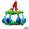
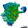
| Title | C1-IgG1 complex on liposomes | |||||||||||||||||||||
 Map data Map data | C1-IgG1 complex observed on liposomes | |||||||||||||||||||||
 Sample Sample |
| |||||||||||||||||||||
| Function / homology |  Function and homology information Function and homology informationcomplement component C1 complex / complement component C1q complex / negative regulation of macrophage differentiation / synapse pruning / negative regulation of granulocyte differentiation / vertebrate eye-specific patterning / complement-mediated synapse pruning / collagen trimer /  complement activation / Classical antibody-mediated complement activation ...complement component C1 complex / complement component C1q complex / negative regulation of macrophage differentiation / synapse pruning / negative regulation of granulocyte differentiation / vertebrate eye-specific patterning / complement-mediated synapse pruning / collagen trimer / complement activation / Classical antibody-mediated complement activation / neuron remodeling / Initial triggering of complement / immunoglobulin complex / complement activation, classical pathway / Regulation of Complement cascade / astrocyte activation / synapse organization / microglial cell activation / cell-cell signaling / amyloid-beta binding / postsynapse / collagen-containing extracellular matrix / blood microparticle / adaptive immune response / immune response / innate immune response / synapse / extracellular space / extracellular region / plasma membrane complement activation / Classical antibody-mediated complement activation ...complement component C1 complex / complement component C1q complex / negative regulation of macrophage differentiation / synapse pruning / negative regulation of granulocyte differentiation / vertebrate eye-specific patterning / complement-mediated synapse pruning / collagen trimer / complement activation / Classical antibody-mediated complement activation / neuron remodeling / Initial triggering of complement / immunoglobulin complex / complement activation, classical pathway / Regulation of Complement cascade / astrocyte activation / synapse organization / microglial cell activation / cell-cell signaling / amyloid-beta binding / postsynapse / collagen-containing extracellular matrix / blood microparticle / adaptive immune response / immune response / innate immune response / synapse / extracellular space / extracellular region / plasma membraneSimilarity search - Function | |||||||||||||||||||||
| Biological species |  Homo sapiens (human) Homo sapiens (human) | |||||||||||||||||||||
| Method | subtomogram averaging / cryo EM / Resolution: 25.0 Å | |||||||||||||||||||||
 Authors Authors | Howes SC / Koning RI / de Jong RN / Beurskens FJ / Koster AJ / Sharp TH | |||||||||||||||||||||
| Funding support |  Netherlands, 6 items Netherlands, 6 items
| |||||||||||||||||||||
 Citation Citation | Journal: Science / Year: 2018 Title: Structures of C1-IgG1 provide insights into how danger pattern recognition activates complement. Authors: Deniz Ugurlar / Stuart C Howes / Bart-Jan de Kreuk / Roman I Koning / Rob N de Jong / Frank J Beurskens / Janine Schuurman / Abraham J Koster / Thomas H Sharp / Paul W H I Parren / Piet Gros / Abstract: Danger patterns on microbes or damaged host cells bind and activate C1, inducing innate immune responses and clearance through the complement cascade. How these patterns trigger complement initiation ...Danger patterns on microbes or damaged host cells bind and activate C1, inducing innate immune responses and clearance through the complement cascade. How these patterns trigger complement initiation remains elusive. Here, we present cryo-electron microscopy analyses of C1 bound to monoclonal antibodies in which we observed heterogeneous structures of single and clustered C1-immunoglobulin G1 (IgG1) hexamer complexes. Distinct C1q binding sites are observed on the two Fc-CH2 domains of each IgG molecule. These are consistent with known interactions and also reveal additional interactions, which are supported by functional IgG1-mutant analysis. Upon antibody binding, the C1q arms condense, inducing rearrangements of the C1rs proteases and tilting C1q's cone-shaped stalk. The data suggest that C1r may activate C1s within single, strained C1 complexes or between neighboring C1 complexes on surfaces. | |||||||||||||||||||||
| History |
|
- Structure visualization
| Movie |
Movie viewer |
|---|---|
| Structure viewer | EM map: SurfViewMolmilJmol/JSmol |
| Supplemental images |
- Downloads & links
-EMDB archive
| Map data | emd_4231.map.gz | 1.7 MB | EMDB map data format | |
|---|---|---|---|---|
| Header (meta data) | emd-4231-v30.xmlemd-4231.xml | 27.3 KB 27.3 KB | Display Display | EMDB header |
| FSC (resolution estimation) | emd_4231_fsc.xml | 3.6 KB | Display | FSC data file |
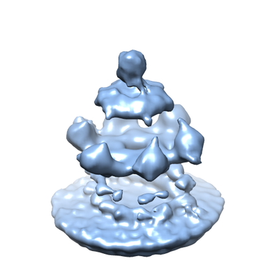
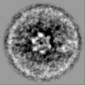
| Images |  emd_4231.png emd_4231.png | 82.6 KB | ||
| Others | emd_4231_additional_1.map.gzemd_4231_additional_2.map.gzemd_4231_additional_3.map.gzemd_4231_additional_4.map.gzemd_4231_half_map_1.map.gzemd_4231_half_map_2.map.gz | 10.8 MB 1.7 MB 10.7 MB 10.7 MB 2.2 MB 2.1 MB | ||
| Archive directory |  http://ftp.pdbj.org/pub/emdb/structures/EMD-4231ftp://ftp.pdbj.org/pub/emdb/structures/EMD-4231 http://ftp.pdbj.org/pub/emdb/structures/EMD-4231ftp://ftp.pdbj.org/pub/emdb/structures/EMD-4231 | HTTPS FTP |
-Related structure data


-Links
| EMDB pages | EMDB (EBI/PDBe) / EMDataResource |
|---|---|
| Related items in Molecule of the Month |


-Map
| File | Download / File: emd_4231.map.gz / Format: CCP4 / Size: 2.3 MB / Type: IMAGE STORED AS FLOATING POINT NUMBER (4 BYTES) | ||||||||||||||||||||||||||||||||||||||||||||||||||||||||||||||||||||
|---|---|---|---|---|---|---|---|---|---|---|---|---|---|---|---|---|---|---|---|---|---|---|---|---|---|---|---|---|---|---|---|---|---|---|---|---|---|---|---|---|---|---|---|---|---|---|---|---|---|---|---|---|---|---|---|---|---|---|---|---|---|---|---|---|---|---|---|---|---|
| Annotation | C1-IgG1 complex observed on liposomes | ||||||||||||||||||||||||||||||||||||||||||||||||||||||||||||||||||||
| Voxel size | X=Y=Z: 5.38 Å | ||||||||||||||||||||||||||||||||||||||||||||||||||||||||||||||||||||
| Density |
| ||||||||||||||||||||||||||||||||||||||||||||||||||||||||||||||||||||
| Symmetry | Space group: 1 | ||||||||||||||||||||||||||||||||||||||||||||||||||||||||||||||||||||
| Details | EMDB XML:
CCP4 map header:
| ||||||||||||||||||||||||||||||||||||||||||||||||||||||||||||||||||||
-Supplemental data
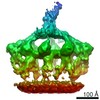
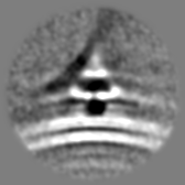
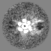
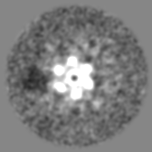
-Additional map: Class 1 of neighboring C1-IgG1 complexes
| File | emd_4231_additional_1.map | ||||||||||||
|---|---|---|---|---|---|---|---|---|---|---|---|---|---|
| Annotation | Class 1 of neighboring C1-IgG1 complexes | ||||||||||||
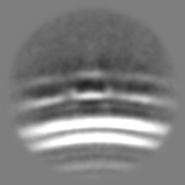
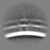
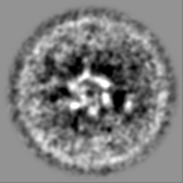
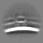
| Projections & Slices |
| ||||||||||||
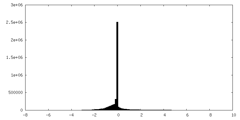
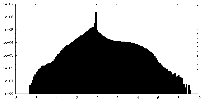
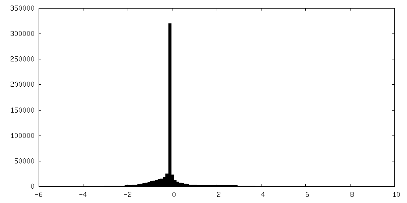
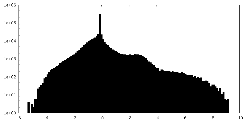
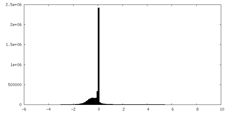
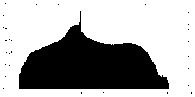
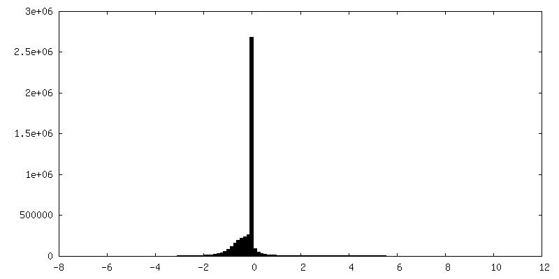
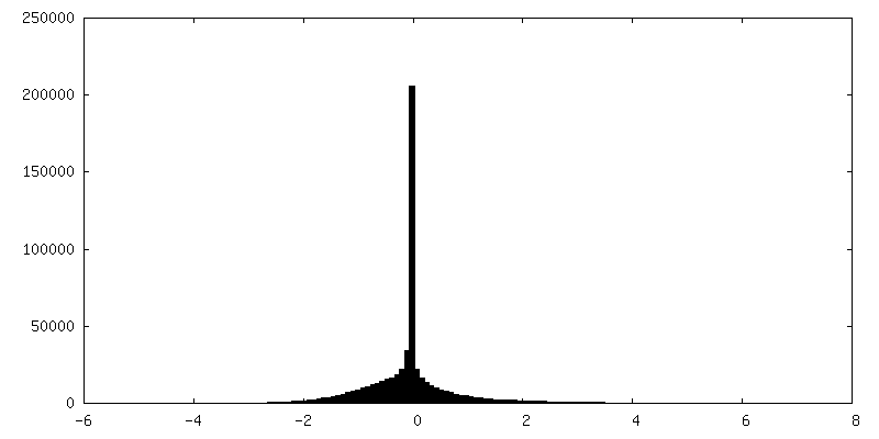
| Density Histograms |
 Z
Z Y
Y X
X


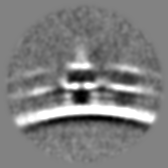
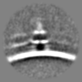
-Additional map: C1-IgG1 complex observed on liposomes refined using mask...
| File | emd_4231_additional_2.map | ||||||||||||
|---|---|---|---|---|---|---|---|---|---|---|---|---|---|
| Annotation | C1-IgG1 complex observed on liposomes refined using mask that excluded the membrane and Fab regions | ||||||||||||
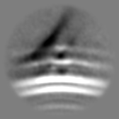
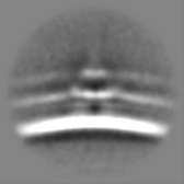
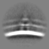
| Projections & Slices |
| ||||||||||||
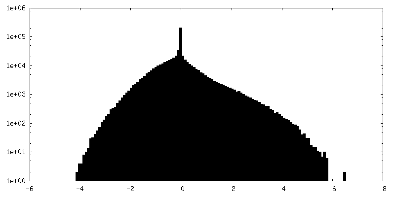
| Density Histograms |


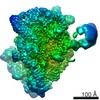
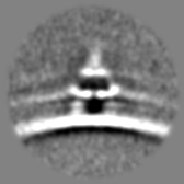
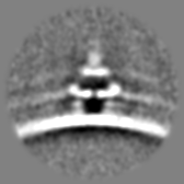
-Additional map: Class 2 of neighboring C1-IgG1 complexes
| File | emd_4231_additional_3.map | ||||||||||||
|---|---|---|---|---|---|---|---|---|---|---|---|---|---|
| Annotation | Class 2 of neighboring C1-IgG1 complexes | ||||||||||||
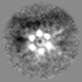
| Projections & Slices |
| ||||||||||||
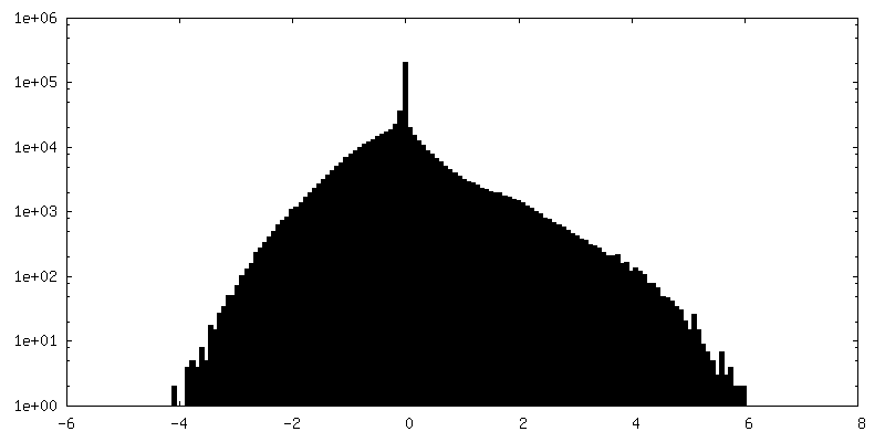
| Density Histograms |
-Additional map: Class 3 of neighboring C1-IgG1 complexes
| File | emd_4231_additional_4.map | ||||||||||||
|---|---|---|---|---|---|---|---|---|---|---|---|---|---|
| Annotation | Class 3 of neighboring C1-IgG1 complexes | ||||||||||||
| Projections & Slices |
| ||||||||||||
| Density Histograms |

-Half map: Half map of C1-IgG1 complex observed on liposomes
| File | emd_4231_half_map_1.map | ||||||||||||
|---|---|---|---|---|---|---|---|---|---|---|---|---|---|
| Annotation | Half map of C1-IgG1 complex observed on liposomes | ||||||||||||
| Projections & Slices |
| ||||||||||||
| Density Histograms |


-Half map: Half map of C1-IgG1 complex observed on liposomes
| File | emd_4231_half_map_2.map | ||||||||||||
|---|---|---|---|---|---|---|---|---|---|---|---|---|---|
| Annotation | Half map of C1-IgG1 complex observed on liposomes | ||||||||||||
| Projections & Slices |
| ||||||||||||
| Density Histograms |


- Sample components
-Entire : C1-IgG1 complex on liposomes
| Entire | Name: C1-IgG1 complex on liposomes |
|---|---|
| Components |
|
-Supramolecule #1: C1-IgG1 complex on liposomes
| Supramolecule | Name: C1-IgG1 complex on liposomes / type: complex / ID: 1 / Parent: 0 Details: Liposomes containing di-nitrophenyl haptens were incubated with monoclonal antibodies and C1 |
|---|---|
| Source (natural) | Organism: Homo sapiens (human) |
| Molecular weight | Theoretical: 700 KDa |
-Supramolecule #2: IgG1 antibodies
| Supramolecule | Name: IgG1 antibodies / type: complex / ID: 2 / Parent: 1 Details: Monoclonal IgG1 recombinantly expressed in HEK293 FreeStyle cells, purified and bound to liposomes. |
|---|---|
| Source (natural) | Organism: Homo sapiens (human) |
| Recombinant expression | Organism: Homo sapiens (human) / Recombinant cell: HEK293 |
-Supramolecule #3: C1 complex
| Supramolecule | Name: C1 complex / type: complex / ID: 3 / Parent: 1 Details: C1 complex purified from human serum containing C1q, C1r, and C1s. Map from focused alignment and classification. |
|---|---|
| Source (natural) | Organism: Homo sapiens (human) |
-Experimental details
-Structure determination
| Method | cryo EM |
|---|---|
 Processing Processing | subtomogram averaging |
| Aggregation state | particle |
-Sample preparation
| Concentration | 1.3 mg/mL | ||||||||||||
|---|---|---|---|---|---|---|---|---|---|---|---|---|---|
| Buffer | pH: 8 Component:
| ||||||||||||
| Grid | Model: Quantifoil R2/1 / Pretreatment - Type: GLOW DISCHARGE / Pretreatment - Atmosphere: AIR | ||||||||||||
| Vitrification | Cryogen name: ETHANE / Chamber humidity: 96 % / Chamber temperature: 294 K / Instrument: LEICA EM GP Details: 3 microlitres applied and incubated for 30 seconds, blot for 1 second before plunging. | ||||||||||||
| Details | Extruded liposomes were incubated with IgG1 and C1. Gold fiducials were added just before applying to grids. |
- Electron microscopy
| Microscope | FEI TITAN KRIOS |
|---|---|
| Electron beam | Acceleration voltage: 300 kV / Electron source: FIELD EMISSION GUN |
| Electron optics | C2 aperture diameter: 70.0 µm / Illumination mode: FLOOD BEAM / Imaging mode: BRIGHT FIELDBright-field microscopy / Cs: 2.7 mm / Nominal defocus min: 0.3 µm / Nominal magnification: 53000 |
| Specialist optics | Phase plate: VOLTA PHASE PLATE / Energy filter - Name: GIF Quantum LS |
| Sample stage | Specimen holder model: FEI TITAN KRIOS AUTOGRID HOLDER / Cooling holder cryogen: NITROGEN |
| Image recording | Film or detector model: GATAN K2 SUMMIT (4k x 4k) / Detector mode: COUNTING / Digitization - Dimensions - Width: 3710 pixel / Digitization - Dimensions - Height: 3838 pixel / Digitization - Sampling interval: 5.0 µm / Digitization - Frames/image: 1-6 / Average electron dose: 1.2 e/Å2 |
| Experimental equipment |  Model: Titan Krios / Image courtesy: FEI Company |
-Image processing
| Extraction | Number tomograms: 34 / Number images used: 51274 / Method: Manually annotate liposome surfaces / Software - Name: Dynamo (ver. 1.1.291) Details: Equally spaced sub-volumes were extracted from the surfaces of liposomes |
|---|---|
| Final 3D classification | Software - Name: Dynamo (ver. 1.1.291) |
| Final angle assignment | Type: OTHER / Software - Name: Dynamo (ver. 1.1.291) Details: Volume matching after filtering reference volume to angular range present in sub-volume |
| Final reconstruction | Applied symmetry - Point group: C1 (asymmetric) / Algorithm: BACK PROJECTION / Resolution.type: BY AUTHOR / Resolution: 25.0 Å / Resolution method: FSC 0.143 CUT-OFF / Software - Name: Dynamo / Number subtomograms used: 1660 |
| FSC plot (resolution estimation) | 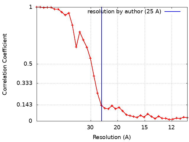 |
