ムービー
ムービー コントローラー


+ データを開く

- 基本情報
基本情報
| 登録情報 | データベース: EMDB / ID: EMD-1074 | |||||||||
|---|---|---|---|---|---|---|---|---|---|---|
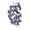
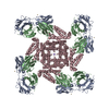
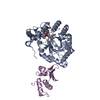
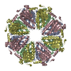
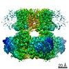
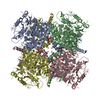
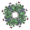
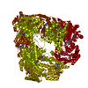
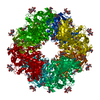
| タイトル | Three-dimensional structure of I(to); Kv4.2-KChIP2 ion channels by electron microscopy at 21 Angstrom resolution. | |||||||||
 マップデータ マップデータ | This is the 3D map of the Kv4.2-KChIP complex | |||||||||
 試料 試料 |
| |||||||||
| 生物種 |   Homo sapiens (ヒト) Homo sapiens (ヒト) | |||||||||
| 手法 | 単粒子再構成法 / ネガティブ染色法 / 解像度: 21.0 Å | |||||||||
 データ登録者 データ登録者 | Kim LA / Furst J / Gutierrez D / Butler MH / Xu S / Goldstein SAN / Grigorieff N | |||||||||
 引用 引用 | ジャーナル: Neuron / 年: 2004 タイトル: Three-dimensional structure of I(to); Kv4.2-KChIP2 ion channels by electron microscopy at 21 Angstrom resolution. 著者: Leo A Kim / Johannes Furst / David Gutierrez / Margaret H Butler / Shuhua Xu / Steve A N Goldstein / Nikolaus Grigorieff /  要旨: Regulatory KChIP2 subunits assemble with pore-forming Kv4.2 subunits in 4:4 complexes to produce native voltage-gated potassium (Kv) channels like cardiac I(to) and neuronal I(A) subtypes. Here, ...Regulatory KChIP2 subunits assemble with pore-forming Kv4.2 subunits in 4:4 complexes to produce native voltage-gated potassium (Kv) channels like cardiac I(to) and neuronal I(A) subtypes. Here, negative stain electron microscopy (EM) and single particle averaging reveal KChIP2 to create a novel approximately 35 x 115 x 115 Angstrom, intracellular fenestrated rotunda: four peripheral columns that extend down from the membrane-embedded portion of the channel to enclose the Kv4.2 "hanging gondola" (a platform held beneath the transmembrane conduction pore by four internal columns). To reach the pore from the cytosol, ions traverse one of four external fenestrae to enter the rotundal vestibule and then cross one of four internal windows in the gondola. | |||||||||
| 履歴 |
|
- 構造の表示
構造の表示
| ムービー |
 ムービービューア ムービービューア |
|---|---|
| 構造ビューア | EMマップ: SurfViewMolmilJmol/JSmol |
| 添付画像 |

- ダウンロードとリンク
-EMDBアーカイブ
| マップデータ | emd_1074.map.gz | 2.6 MB | EMDBマップデータ形式 | |
|---|---|---|---|---|
| ヘッダ (付随情報) | emd-1074-v30.xmlemd-1074.xml | 11.7 KB 11.7 KB | 表示 表示 | EMDBヘッダ |
| 画像 |  1074.gif 1074.gif | 26.5 KB | ||
| アーカイブディレクトリ |  http://ftp.pdbj.org/pub/emdb/structures/EMD-1074ftp://ftp.pdbj.org/pub/emdb/structures/EMD-1074 http://ftp.pdbj.org/pub/emdb/structures/EMD-1074ftp://ftp.pdbj.org/pub/emdb/structures/EMD-1074 | HTTPS FTP |
-関連構造データ
-リンク
| EMDBのページ | EMDB (EBI/PDBe) / EMDataResource |
|---|
-マップ
| ファイル | ダウンロード / ファイル: emd_1074.map.gz / 形式: CCP4 / 大きさ: 2.7 MB / タイプ: IMAGE STORED AS FLOATING POINT NUMBER (4 BYTES) | ||||||||||||||||||||||||||||||||||||||||||||||||||||||||||||||||||||
|---|---|---|---|---|---|---|---|---|---|---|---|---|---|---|---|---|---|---|---|---|---|---|---|---|---|---|---|---|---|---|---|---|---|---|---|---|---|---|---|---|---|---|---|---|---|---|---|---|---|---|---|---|---|---|---|---|---|---|---|---|---|---|---|---|---|---|---|---|---|
| 注釈 | This is the 3D map of the Kv4.2-KChIP complex | ||||||||||||||||||||||||||||||||||||||||||||||||||||||||||||||||||||
| ボクセルのサイズ | X=Y=Z: 4.67 Å | ||||||||||||||||||||||||||||||||||||||||||||||||||||||||||||||||||||
| 密度 |
| ||||||||||||||||||||||||||||||||||||||||||||||||||||||||||||||||||||
| 対称性 | 空間群: 1 | ||||||||||||||||||||||||||||||||||||||||||||||||||||||||||||||||||||
| 詳細 | EMDB XML:
CCP4マップ ヘッダ情報:
| ||||||||||||||||||||||||||||||||||||||||||||||||||||||||||||||||||||
-添付データ
- 試料の構成要素
-全体 : Human Kv4.2-KChIP2 Ion Channel Complex
| 全体 | 名称: Human Kv4.2-KChIP2 Ion Channel Complex |
|---|---|
| 要素 |
|
-超分子 #1000: Human Kv4.2-KChIP2 Ion Channel Complex
| 超分子 | 名称: Human Kv4.2-KChIP2 Ion Channel Complex / タイプ: sample / ID: 1000 詳細: The sample was solubilised in 0.7% CHAPS and was monodisperse 集合状態: 4 Kv4.2 subunits bind to 4 KChIP2 subunits / Number unique components: 2 |
|---|---|
| 分子量 | 実験値: 440 KDa / 理論値: 400 KDa / 手法: SDS-PAGE |
-分子 #1: Kv4.2
| 分子 | 名称: Kv4.2 / タイプ: protein_or_peptide / ID: 1 / 詳細: 4 monomers form a membrane-embedded channel / コピー数: 4 / 集合状態: tetramer / 組換発現: Yes |
|---|---|
| 由来(天然) | 生物種: Homo sapiens (ヒト) / 別称: Human / 細胞中の位置: Cell membrane |
| 分子量 | 実験値: 72 MDa / 理論値: 75 MDa |
| 組換発現 | 生物種: COS7 / 組換プラスミド: pRAT |
-分子 #2: KChIP2
| 分子 | 名称: KChIP2 / タイプ: ligand / ID: 2 詳細: 4 monomers bind on the cytoplasmic side of the channel コピー数: 4 / 集合状態: Monomer / 組換発現: Yes |
|---|---|
| 分子量 | 実験値: 28 MDa / 理論値: 35 MDa |
| 組換発現 | 生物種: COS7 / 組換プラスミド: pRAT |
-実験情報
-構造解析
| 手法 | ネガティブ染色法 |
|---|---|
 解析 解析 | 単粒子再構成法 |
| 試料の集合状態 | particle |
-試料調製
| 濃度 | 0.1 mg/mL |
|---|---|
| 緩衝液 | pH: 7.4 詳細: 0.7% CHAPS, 100 mM NaCl, 40 mM KCl, 0.01 mM leupeptin/pepstatin, 1 mM EDTA, 20 mM HEPES-KOH (pH 7.4) |
| 染色 | タイプ: NEGATIVE 詳細: Grids with adsorbed protein were washed 3 times in detergent-free buffer and then floated on 1% uranyl acetate for 10 seconds |
| グリッド | 詳細: 400 mesh copper grid |
| 凍結 | 凍結剤: NONE |
- 電子顕微鏡法
| 顕微鏡 | FEI/PHILIPS CM120T |
|---|---|
| 電子線 | 加速電圧: 120 kV / 電子線源: LAB6 |
| 電子光学系 | 照射モード: FLOOD BEAM / 撮影モード: BRIGHT FIELDBright-field microscopy / Cs: 2.0 mm / 最大 デフォーカス(公称値): 1.5 µm / 最小 デフォーカス(公称値): 1.5 µm / 倍率(公称値): 60000 |
| 試料ステージ | 試料ホルダー: Eucentric, room temperature / 試料ホルダーモデル: OTHER |
| 温度 | 平均: 300 K |
| アライメント法 | Legacy - 非点収差: carbon film grain |
| 日付 | 2003年6月1日 |
| 撮影 | カテゴリ: FILM / フィルム・検出器のモデル: KODAK SO-163 FILM / デジタル化 - スキャナー: ZEISS SCAI / デジタル化 - サンプリング間隔: 4.67 µm / 実像数: 63 / 平均電子線量: 10 e/Å2 詳細: Images were scanned at 7 microns resolution and 4 x 4 pixels were averaged Od range: 1.4 / ビット/ピクセル: 8 |
| Tilt angle min | 0 |
| Tilt angle max | 0 |
-画像解析
| CTF補正 | 詳細: Each particle |
|---|---|
| 最終 角度割当 | 詳細: theta 90 degrees, phi 90 degrees |
| 最終 再構成 | 想定した対称性 - 点群: C4 (4回回転対称) / アルゴリズム: OTHER / 解像度のタイプ: BY AUTHOR / 解像度: 21.0 Å / 解像度の算出法: FSC 0.5 CUT-OFF / ソフトウェア - 名称: IMAGIC, FREALIGN / 詳細: FREALIGN reconstruction in Fourier space / 使用した粒子像数: 8164 |
| 詳細 | particles were selected manually |