 Movie
Movie Controller

[English] 日本語
 Yorodumi
- PDB-1orq: X-ray structure of a voltage-dependent potassium channel in compl... -
+ Open data

- Basic information
| Entry | Database: PDB / ID: 1orq | ||||||
|---|---|---|---|---|---|---|---|
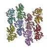
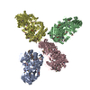
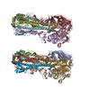
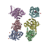
| Title | X-ray structure of a voltage-dependent potassium channel in complex with an Fab | ||||||
 Components Components |
| ||||||
 Keywords Keywords |  MEMBRANE PROTEIN / potassium channel / voltage-dependent / KvAP / Fab complex MEMBRANE PROTEIN / potassium channel / voltage-dependent / KvAP / Fab complex | ||||||
| Function / homology |  Function and homology information Function and homology informationpositive regulation of B cell activation / early endosome to late endosome transport / humoral immune response mediated by circulating immunoglobulin / phagocytosis, recognition / positive regulation of type IIa hypersensitivity / regulation of proteolysis / positive regulation of type I hypersensitivity / antibody-dependent cellular cytotoxicity / Fc-gamma receptor I complex binding / monoatomic ion channel complex ...positive regulation of B cell activation / early endosome to late endosome transport / humoral immune response mediated by circulating immunoglobulin / phagocytosis, recognition / positive regulation of type IIa hypersensitivity / regulation of proteolysis / positive regulation of type I hypersensitivity / antibody-dependent cellular cytotoxicity / Fc-gamma receptor I complex binding / monoatomic ion channel complex / phagocytosis, engulfment / endosome to lysosome transport / positive regulation of endocytosis / immunoglobulin complex, circulating / IgG immunoglobulin complex / immunoglobulin receptor binding / potassium channel activity / immunoglobulin mediated immune response / antigen processing and presentation / positive regulation of phagocytosis / complement activation, classical pathway / antigen binding / multivesicular body / B cell differentiation / response to bacterium / positive regulation of immune response / antibacterial humoral response / extracellular space / extracellular region / identical protein binding / plasma membrane / cytosolSimilarity search - Function | ||||||
| Biological species |   Aeropyrum pernix (archaea) Aeropyrum pernix (archaea) Mus musculus (house mouse) Mus musculus (house mouse) | ||||||
| Method | X-RAY DIFFRACTION / SYNCHROTRON / MOLECULAR REPLACEMENT / Resolution: 3.2 Å | ||||||
 Authors Authors | Jiang, Y. / Lee, A. / Chen, J. / Ruta, V. / Cadene, M. / Chait, B.T. / MacKinnon, R. | ||||||
 Citation Citation | Journal: Nature / Year: 2003 Title: X-ray structure of a voltage-dependent K+ channel Authors: Jiang, Y. / Lee, A. / Chen, J. / Ruta, V. / Cadene, M. / Chait, B.T. / MacKinnon, R. | ||||||
| History |
|
- Structure visualization
| Structure viewer | Molecule: MolmilJmol/JSmol |
|---|
- Downloads & links
-Download
| PDBx/mmCIF format | 1orq.cif.gz | 141 KB | Display | PDBx/mmCIF format |
|---|---|---|---|---|
| PDB format | pdb1orq.ent.gz | 109.1 KB | Display | PDB format |
| PDBx/mmJSON format | 1orq.json.gz | Tree view | PDBx/mmJSON format | |
| Others |  Other downloads Other downloads |
-Validation report
| Arichive directory | https://data.pdbj.org/pub/pdb/validation_reports/or/1orqftp://data.pdbj.org/pub/pdb/validation_reports/or/1orq | HTTPS FTP |
|---|
-Related structure data
| Related structure data | 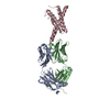 1orsC 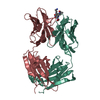 1bafS C: citing same article ( S: Starting model for refinement |
|---|---|
| Similar structure data |


-Links
 PDBj
PDBj


- Assembly
| Deposited unit | 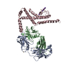
| ||||||||||||||||||||||||
|---|---|---|---|---|---|---|---|---|---|---|---|---|---|---|---|---|---|---|---|---|---|---|---|---|---|
| 1 | 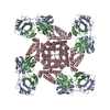
| ||||||||||||||||||||||||
| 2 | x 8
| ||||||||||||||||||||||||
| Unit cell |
| ||||||||||||||||||||||||
| Components on special symmetry positions |
| ||||||||||||||||||||||||
| Details | The channel functions as tetramer. The remainder of the biological assembly is generated by the four fold axis: -x -y z, -y x z, y -x z. |
-Components
-Protein , 1 types, 1 molecules C
| #3: Protein | Mass: 24271.910 Da / Num. of mol.: 1 / Fragment: KvAP / Mutation: Y46C Source method: isolated from a genetically manipulated source Source: (gene. exp.) Aeropyrum pernix (archaea) / Plasmid: pQE60 / Production host:  Escherichia coli (E. coli) / References: UniProt: Q9YDF8 Escherichia coli (E. coli) / References: UniProt: Q9YDF8 |
|---|
-Antibody , 2 types, 2 molecules AB
| #1: Antibody | Mass: 23527.826 Da / Num. of mol.: 1 / Source method: isolated from a natural source / Details: mouse hybridoma / Source: (natural) Mus musculus (house mouse) / References: UniProt: P01837 |
|---|---|
| #2: Antibody | Mass: 23781.516 Da / Num. of mol.: 1 / Source method: isolated from a natural source / Details: mouse hybridoma / Source: (natural) Mus musculus (house mouse) / References: UniProt: P01865 |
-Non-polymers , 3 types, 19 molecules 


| #4: Chemical | ChemComp-CD /  Mass: 112.411 Da / Num. of mol.: 7 / Source method: obtained synthetically / Formula: Cd Mass: 112.411 Da / Num. of mol.: 7 / Source method: obtained synthetically / Formula: Cd#5: Chemical | ChemComp-K /  Mass: 39.098 Da / Num. of mol.: 6 / Source method: obtained synthetically / Formula: K Mass: 39.098 Da / Num. of mol.: 6 / Source method: obtained synthetically / Formula: K#6: Water | ChemComp-HOH / | WaterMass: 18.015 Da / Num. of mol.: 6 / Source method: isolated from a natural source / Formula: H2O |
|---|
-Experimental details
-Experiment
| Experiment | Method: X-RAY DIFFRACTION / Number of used crystals: 1 |
|---|
- Sample preparation
| Crystal | Density Matthews: 4.77 Å3/Da / Density % sol: 73.99 % | ||||||||||||||||||||||||||||||||||||||||||||||||||||||||
|---|---|---|---|---|---|---|---|---|---|---|---|---|---|---|---|---|---|---|---|---|---|---|---|---|---|---|---|---|---|---|---|---|---|---|---|---|---|---|---|---|---|---|---|---|---|---|---|---|---|---|---|---|---|---|---|---|---|
| Crystal grow | Temperature: 293 K / Method: vapor diffusion, sitting drop / pH: 5 Details: PEG400, Cadmium chloride, Sodium acetate, pH 5.0, VAPOR DIFFUSION, SITTING DROP, temperature 293K | ||||||||||||||||||||||||||||||||||||||||||||||||||||||||
| Crystal grow | *PLUS Temperature: 20 ℃ / pH: 8 | ||||||||||||||||||||||||||||||||||||||||||||||||||||||||
| Components of the solutions | *PLUS
|
-Data collection
| Diffraction | Mean temperature: 100 K |
|---|---|
| Diffraction source | Source: SYNCHROTRON / Site: CHESS  / Beamline: A1 / Wavelength: 0.937 Å / Beamline: A1 / Wavelength: 0.937 Å |
| Detector | Type: ADSC QUANTUM 4 / Detector: CCD / Date: Oct 5, 2002 |
| Radiation | Protocol: SINGLE WAVELENGTH / Monochromatic (M) / Laue (L): M / Scattering type: x-ray |
| Radiation wavelength | Wavelength: 0.937 Å / Relative weight: 1 |
| Reflection | Resolution: 3.2→30 Å / Num. all: 22626 / Num. obs: 22476 / % possible obs: 98.5 % / Observed criterion σ(I): 0 / Redundancy: 5.5 % / Rsym value: 0.08 / Net I/σ(I): 18 |
| Reflection shell | Resolution: 3.2→3.31 Å / Redundancy: 3 % / Mean I/σ(I) obs: 2 / Num. unique all: 22501 / Rsym value: 0.424 / % possible all: 95.2 |
| Reflection | *PLUS Lowest resolution: 30 Å / Rmerge(I) obs: 0.08 |
| Reflection shell | *PLUS % possible obs: 95.2 % / Rmerge(I) obs: 0.424 / Mean I/σ(I) obs: 2.2 |
- Processing
| Software |
| |||||||||||||||||||||||||
|---|---|---|---|---|---|---|---|---|---|---|---|---|---|---|---|---|---|---|---|---|---|---|---|---|---|---|
| Refinement | Method to determine structure: MOLECULAR REPLACEMENT Starting model: pdb code 1BAF Resolution: 3.2→29.95 Å / Rfactor Rfree error: 0.009 / Isotropic thermal model: RESTRAINED / Cross valid method: THROUGHOUT / σ(F): 0 / Stereochemistry target values: Engh & Huber
| |||||||||||||||||||||||||
| Solvent computation | Solvent model: FLAT MODEL / Bsol: 26.7975 Å2 / ksol: 0.252452 e/Å3 | |||||||||||||||||||||||||
| Displacement parameters | Biso mean: 73.4 Å2
| |||||||||||||||||||||||||
| Refine analyze | Luzzati coordinate error free: 0.49 Å / Luzzati sigma a free: 0.89 Å | |||||||||||||||||||||||||
| Refinement step | Cycle: LAST / Resolution: 3.2→29.95 Å
| |||||||||||||||||||||||||
| Refine LS restraints |
| |||||||||||||||||||||||||
| LS refinement shell | Resolution: 3.2→3.4 Å / Rfactor Rfree error: 0.029 / Total num. of bins used: 6
| |||||||||||||||||||||||||
| Xplor file |
| |||||||||||||||||||||||||
| Software | *PLUS Version: 1 / Classification: refinement | |||||||||||||||||||||||||
| Refinement | *PLUS σ(F): 0 / % reflection Rfree: 5.1 % | |||||||||||||||||||||||||
| Solvent computation | *PLUS | |||||||||||||||||||||||||
| Displacement parameters | *PLUS Biso mean: 73.4 Å2 | |||||||||||||||||||||||||
| Refine LS restraints | *PLUS
| |||||||||||||||||||||||||
| LS refinement shell | *PLUS Rfactor Rfree: 0.383 / % reflection Rfree: 5 % / Rfactor Rwork: 0.339 |
