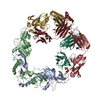
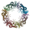
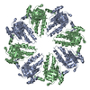
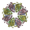
- PDB-4hyy: Filament of octameric rings of DMC1 recombinase from Homo sapiens -
+
Open data
ID or keywords:
Loading...
-
Basic information
Entry
Database: PDB / ID: 4hyy
Title
Filament of octameric rings of DMC1 recombinase from Homo sapiens
Components
Meiotic recombination protein DMC1/LIM15 homologGenetic recombination
Keywords
RECOMBINATION / RecA homolog / DNA strand exchange / DNA / nucleus
Function / homology
Function and homology information
female gamete generation / chromosome organization involved in meiotic cell cycle / DNA recombinase assembly / homologous chromosome pairing at meiosis / double-strand break repair involved in meiotic recombination / mitotic recombination / DNA strand invasion / DNA strand exchange activity / lateral element / oocyte maturation ...female gamete generation / chromosome organization involved in meiotic cell cycle / DNA recombinase assembly / homologous chromosome pairing at meiosis / double-strand break repair involved in meiotic recombination / mitotic recombination / DNA strand invasion / DNA strand exchange activity / lateral element / oocyte maturation / reciprocal meiotic recombination / ATP-dependent DNA damage sensor activity / male meiosis I / spermatid development / ATP-dependent activity, acting on DNA / ovarian follicle development / meiotic cell cycle / condensed nuclear chromosome / Meiotic recombination / single-stranded DNA binding / site of double-strand break / chromosome / double-stranded DNA binding / spermatogenesis / chromosome, telomeric region / ATP hydrolysis activity / DNA binding / ATP binding / identical protein binding / nucleus Similarity search - Function
Meiotic recombination protein Dmc1 / DNA recombination and repair protein, RecA-like / DNA recombination and repair protein Rad51-like, C-terminal / Rad51 / DNA recombination and repair protein RecA, monomer-monomer interface / RecA family profile 2. / DNA recombination and repair protein RecA-like, ATP-binding domain / RecA family profile 1. / DNA repair Rad51/transcription factor NusA, alpha-helical / Helix-hairpin-helix domain ...Meiotic recombination protein Dmc1 / DNA recombination and repair protein, RecA-like / DNA recombination and repair protein Rad51-like, C-terminal / Rad51 / DNA recombination and repair protein RecA, monomer-monomer interface / RecA family profile 2. / DNA recombination and repair protein RecA-like, ATP-binding domain / RecA family profile 1. / DNA repair Rad51/transcription factor NusA, alpha-helical / Helix-hairpin-helix domain / P-loop containing nucleotide triphosphate hydrolases / ATPases associated with a variety of cellular activities / AAA+ ATPase domain / P-loop containing nucleoside triphosphate hydrolase / Rossmann fold / 3-Layer(aba) Sandwich / Alpha Beta Similarity search - Domain/homology
In the structure databanks used in Yorodumi, some data are registered as the other names, "COVID-19 virus" and "2019-nCoV". Here are the details of the virus and the list of structure data.
Jan 31, 2019. EMDB accession codes are about to change! (news from PDBe EMDB page)
EMDB accession codes are about to change! (news from PDBe EMDB page)
The allocation of 4 digits for EMDB accession codes will soon come to an end. Whilst these codes will remain in use, new EMDB accession codes will include an additional digit and will expand incrementally as the available range of codes is exhausted. The current 4-digit format prefixed with “EMD-” (i.e. EMD-XXXX) will advance to a 5-digit format (i.e. EMD-XXXXX), and so on. It is currently estimated that the 4-digit codes will be depleted around Spring 2019, at which point the 5-digit format will come into force.
The EM Navigator/Yorodumi systems omit the EMD- prefix.
Related info.:Q: What is EMD? / ID/Accession-code notation in Yorodumi/EM Navigator
Yorodumi is a browser for structure data from EMDB, PDB, SASBDB, etc.
This page is also the successor to EM Navigator detail page, and also detail information page/front-end page for Omokage search.
The word "yorodu" (or yorozu) is an old Japanese word meaning "ten thousand". "mi" (miru) is to see.
Related info.:EMDB / PDB / SASBDB / Comparison of 3 databanks / Yorodumi Search / Aug 31, 2016. New EM Navigator & Yorodumi / Yorodumi Papers / Jmol/JSmol / Function and homology information / Changes in new EM Navigator and Yorodumi
 Movie
Movie Controller

 Yorodumi
 Open data
 Basic information
 Components
 Genetic recombination
  Keywords
 Function and homology information
 Authors
 Citation
 Structure visualization
 Downloads & links
 Other downloads
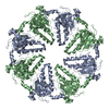


 PDBj
PDBj


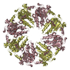
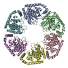
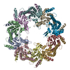
 Assembly
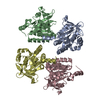


 Mass: 18.015 Da / Num. of mol.: 96 / Source method: isolated from a natural source / Formula: H2O
 Sample preparation
 / Beamline: 08ID-1 / Wavelength: 0.98 Å
 Processing