 Movie
Movie Controller


+ Open data

- Basic information
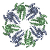
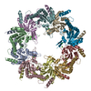
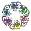
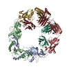
| Entry | Database: PDB / ID: 6r3p | |||||||||
|---|---|---|---|---|---|---|---|---|---|---|
| Title | Crystal structure of human DMC1 ATPase domain | |||||||||
 Components Components | Meiotic recombination protein DMC1/LIM15 homolog Genetic recombination Genetic recombination | |||||||||
 Keywords Keywords | RECOMBINATION / Recombinase / Homologous recombination / Strand invasion / Meiosis | |||||||||
| Function / homology |  Function and homology information Function and homology informationfemale gamete generation / chromosome organization involved in meiotic cell cycle / DNA recombinase assembly / homologous chromosome pairing at meiosis / double-strand break repair involved in meiotic recombination / mitotic recombination / DNA strand invasion / DNA strand exchange activity / lateral element / oocyte maturation ...female gamete generation / chromosome organization involved in meiotic cell cycle / DNA recombinase assembly / homologous chromosome pairing at meiosis / double-strand break repair involved in meiotic recombination / mitotic recombination / DNA strand invasion / DNA strand exchange activity / lateral element / oocyte maturation / reciprocal meiotic recombination / ATP-dependent DNA damage sensor activity / male meiosis I / spermatid development / ATP-dependent activity, acting on DNA / ovarian follicle development / meiotic cell cycle / condensed nuclear chromosome / Meiotic recombination / single-stranded DNA binding / site of double-strand break / chromosome / double-stranded DNA binding / spermatogenesis / chromosome, telomeric region / ATP hydrolysis activity / DNA binding / ATP binding / identical protein binding / nucleusSimilarity search - Function | |||||||||
| Biological species |  Homo sapiens (human) Homo sapiens (human) | |||||||||
| Method | X-RAY DIFFRACTION / SYNCHROTRON / MOLECULAR REPLACEMENT / Resolution: 2.05 Å | |||||||||
 Authors Authors | Dunce, J.M. / Davies, O.R. | |||||||||
| Funding support |  United Kingdom, 2items United Kingdom, 2items
| |||||||||
 Citation Citation | Journal: To Be Published Title: Crystal structure of human DMC1 ATPase domain Authors: Dunce, J.M. / Davies, O.R. | |||||||||
| History |
|
- Structure visualization
| Structure viewer | Molecule: MolmilJmol/JSmol |
|---|
- Downloads & links
-Download
| PDBx/mmCIF format | 6r3p.cif.gz | 571.4 KB | Display | PDBx/mmCIF format |
|---|---|---|---|---|
| PDB format | pdb6r3p.ent.gz | 486.4 KB | Display | PDB format |
| PDBx/mmJSON format | 6r3p.json.gz | Tree view | PDBx/mmJSON format | |
| Others |  Other downloads Other downloads |
-Validation report
| Arichive directory | https://data.pdbj.org/pub/pdb/validation_reports/r3/6r3pftp://data.pdbj.org/pub/pdb/validation_reports/r3/6r3p | HTTPS FTP |
|---|
-Related structure data
| Related structure data |  4hyyS S: Starting model for refinement |
|---|---|
| Similar structure data |


-Links
 PDBj
PDBj


- Assembly
| Deposited unit | 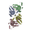
| ||||||||
|---|---|---|---|---|---|---|---|---|---|
| 1 | 
| ||||||||
| 2 | 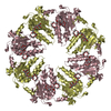
| ||||||||
| Unit cell |
|
-Components
-Protein , 1 types, 4 molecules ABCD
| #1: Protein | Genetic recombination Mass: 28868.811 Da / Num. of mol.: 4 Source method: isolated from a genetically manipulated source Source: (gene. exp.) Homo sapiens (human) / Gene: DMC1, DMC1H, LIM15 / Plasmid: pHAT4 / Production host:  Escherichia coli BL21(DE3) (bacteria) / References: UniProt: Q14565 Escherichia coli BL21(DE3) (bacteria) / References: UniProt: Q14565 |
|---|
-Non-polymers , 6 types, 301 molecules 


| #2: Chemical | Ethylene glycol Mass: 62.068 Da / Num. of mol.: 2 / Source method: obtained synthetically / Formula: C2H6O2 Mass: 62.068 Da / Num. of mol.: 2 / Source method: obtained synthetically / Formula: C2H6O2#3: Chemical | Diethylene glycol Mass: 106.120 Da / Num. of mol.: 2 / Source method: obtained synthetically / Formula: C4H10O3 Mass: 106.120 Da / Num. of mol.: 2 / Source method: obtained synthetically / Formula: C4H10O3#4: Chemical | ChemComp-PG4 / | Polyethylene glycol Mass: 194.226 Da / Num. of mol.: 1 / Source method: obtained synthetically / Formula: C8H18O5 / Comment: precipitant*YM Mass: 194.226 Da / Num. of mol.: 1 / Source method: obtained synthetically / Formula: C8H18O5 / Comment: precipitant*YM#5: Chemical | Polyethylene glycol Mass: 282.331 Da / Num. of mol.: 2 / Source method: obtained synthetically / Formula: C12H26O7 / Comment: precipitant*YM Mass: 282.331 Da / Num. of mol.: 2 / Source method: obtained synthetically / Formula: C12H26O7 / Comment: precipitant*YM#6: Chemical | ChemComp-1PE / | Polyethylene glycol Mass: 238.278 Da / Num. of mol.: 1 / Source method: obtained synthetically / Formula: C10H22O6 / Comment: precipitant*YM Mass: 238.278 Da / Num. of mol.: 1 / Source method: obtained synthetically / Formula: C10H22O6 / Comment: precipitant*YM#7: Water | ChemComp-HOH / | WaterMass: 18.015 Da / Num. of mol.: 293 / Source method: isolated from a natural source / Formula: H2O |
|---|
-Experimental details
-Experiment
| Experiment | Method: X-RAY DIFFRACTION / Number of used crystals: 1 |
|---|
- Sample preparation
| Crystal | Density Matthews: 2.96 Å3/Da / Density % sol: 58.43 % |
|---|---|
| Crystal grow | Temperature: 293 K / Method: vapor diffusion, hanging drop / pH: 7.2 Details: 50 mM HEPES-NaOH pH 7.2, 50 mM MgCl2, 500 mM NaCl, 7.5 % PEG3350 |
-Data collection
| Diffraction | Mean temperature: 100 K / Serial crystal experiment: N |
|---|---|
| Diffraction source | Source: SYNCHROTRON / Site: Diamond / Beamline: I04-1 / Wavelength: 0.9159 Å |
| Detector | Type: DECTRIS PILATUS 6M / Detector: PIXEL / Date: Apr 15, 2018 |
| Radiation | Protocol: SINGLE WAVELENGTH / Monochromatic (M) / Laue (L): M / Scattering type: x-ray |
| Radiation wavelength | Wavelength: 0.9159 Å / Relative weight: 1 |
| Reflection | Resolution: 2.05→47.49 Å / Num. obs: 86315 / % possible obs: 100 % / Redundancy: 15 % / Biso Wilson estimate: 58.74 Å2 / CC1/2: 1 / Rpim(I) all: 0.034 / Rrim(I) all: 0.096 / Net I/σ(I): 19.5 |
| Reflection shell | Resolution: 2.05→2.09 Å / Redundancy: 15.5 % / Mean I/σ(I) obs: 1.6 / Num. unique obs: 4545 / CC1/2: 0.628 / Rpim(I) all: 0.72 / Rrim(I) all: 2.038 / % possible all: 100 |
- Processing
| Software |
| |||||||||||||||||||||||||||||||||||||||||||||||||||||||||||||||||||||||||||||||||||||||||||||||||||||||||||||||||||||||||||||||||||||||||||||||||||||||||||||||||||||||||||||||||||||||||||||||||||||||||||||||||||||||||||||||||
|---|---|---|---|---|---|---|---|---|---|---|---|---|---|---|---|---|---|---|---|---|---|---|---|---|---|---|---|---|---|---|---|---|---|---|---|---|---|---|---|---|---|---|---|---|---|---|---|---|---|---|---|---|---|---|---|---|---|---|---|---|---|---|---|---|---|---|---|---|---|---|---|---|---|---|---|---|---|---|---|---|---|---|---|---|---|---|---|---|---|---|---|---|---|---|---|---|---|---|---|---|---|---|---|---|---|---|---|---|---|---|---|---|---|---|---|---|---|---|---|---|---|---|---|---|---|---|---|---|---|---|---|---|---|---|---|---|---|---|---|---|---|---|---|---|---|---|---|---|---|---|---|---|---|---|---|---|---|---|---|---|---|---|---|---|---|---|---|---|---|---|---|---|---|---|---|---|---|---|---|---|---|---|---|---|---|---|---|---|---|---|---|---|---|---|---|---|---|---|---|---|---|---|---|---|---|---|---|---|---|---|---|---|---|---|---|---|---|---|---|---|---|---|---|---|---|---|
| Refinement | Method to determine structure: MOLECULAR REPLACEMENT Starting model: 4HYY Resolution: 2.05→47.011 Å / SU ML: 0.25 / Cross valid method: FREE R-VALUE / σ(F): 0.33 / Phase error: 23.57
| |||||||||||||||||||||||||||||||||||||||||||||||||||||||||||||||||||||||||||||||||||||||||||||||||||||||||||||||||||||||||||||||||||||||||||||||||||||||||||||||||||||||||||||||||||||||||||||||||||||||||||||||||||||||||||||||||
| Solvent computation | Shrinkage radii: 0.9 Å / VDW probe radii: 1.11 Å | |||||||||||||||||||||||||||||||||||||||||||||||||||||||||||||||||||||||||||||||||||||||||||||||||||||||||||||||||||||||||||||||||||||||||||||||||||||||||||||||||||||||||||||||||||||||||||||||||||||||||||||||||||||||||||||||||
| Refinement step | Cycle: LAST / Resolution: 2.05→47.011 Å
| |||||||||||||||||||||||||||||||||||||||||||||||||||||||||||||||||||||||||||||||||||||||||||||||||||||||||||||||||||||||||||||||||||||||||||||||||||||||||||||||||||||||||||||||||||||||||||||||||||||||||||||||||||||||||||||||||
| Refine LS restraints |
| |||||||||||||||||||||||||||||||||||||||||||||||||||||||||||||||||||||||||||||||||||||||||||||||||||||||||||||||||||||||||||||||||||||||||||||||||||||||||||||||||||||||||||||||||||||||||||||||||||||||||||||||||||||||||||||||||
| LS refinement shell |
| |||||||||||||||||||||||||||||||||||||||||||||||||||||||||||||||||||||||||||||||||||||||||||||||||||||||||||||||||||||||||||||||||||||||||||||||||||||||||||||||||||||||||||||||||||||||||||||||||||||||||||||||||||||||||||||||||
| Refinement TLS params. | Method: refined / Refine-ID: X-RAY DIFFRACTION
| |||||||||||||||||||||||||||||||||||||||||||||||||||||||||||||||||||||||||||||||||||||||||||||||||||||||||||||||||||||||||||||||||||||||||||||||||||||||||||||||||||||||||||||||||||||||||||||||||||||||||||||||||||||||||||||||||
| Refinement TLS group |
|
