 Movie
Movie Controller
[English] 日本語
 Yorodumi
- SASDAT6: Integrin beta4, fragment of the cytoplasmic region encompassing t... -
+ Open data

- Basic information
| Entry | Database: SASBDB / ID: SASDAT6 |
|---|---|
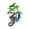
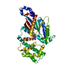
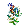
 Sample Sample | Integrin beta4, fragment of the cytoplasmic region encompassing the third and fourth FnIII domains
|
| Function / homology |  Function and homology information Function and homology informationtrophoblast cell migration / Type I hemidesmosome assembly /  hemidesmosome assembly / nail development / hemidesmosome / peripheral nervous system myelin formation / skin morphogenesis / Laminin interactions / filopodium assembly / mesodermal cell differentiation ...trophoblast cell migration / Type I hemidesmosome assembly / hemidesmosome assembly / nail development / hemidesmosome / peripheral nervous system myelin formation / skin morphogenesis / Laminin interactions / filopodium assembly / mesodermal cell differentiation / integrin complex / Assembly of collagen fibrils and other multimeric structures / cell adhesion mediated by integrin / Syndecan interactions / cell leading edge / basement membrane / cell-matrix adhesion / basal plasma membrane / integrin-mediated signaling pathway / G protein-coupled receptor binding / cell motility / autophagy / cell-cell adhesion / response to wounding / integrin binding / cell junction / nuclear membrane / receptor complex / cell adhesion / focal adhesion / nucleolus / cell surface / extracellular exosome / metal ion binding / plasma membrane hemidesmosome assembly / nail development / hemidesmosome / peripheral nervous system myelin formation / skin morphogenesis / Laminin interactions / filopodium assembly / mesodermal cell differentiation ...trophoblast cell migration / Type I hemidesmosome assembly / hemidesmosome assembly / nail development / hemidesmosome / peripheral nervous system myelin formation / skin morphogenesis / Laminin interactions / filopodium assembly / mesodermal cell differentiation / integrin complex / Assembly of collagen fibrils and other multimeric structures / cell adhesion mediated by integrin / Syndecan interactions / cell leading edge / basement membrane / cell-matrix adhesion / basal plasma membrane / integrin-mediated signaling pathway / G protein-coupled receptor binding / cell motility / autophagy / cell-cell adhesion / response to wounding / integrin binding / cell junction / nuclear membrane / receptor complex / cell adhesion / focal adhesion / nucleolus / cell surface / extracellular exosome / metal ion binding / plasma membraneSimilarity search - Function |
| Biological species |  Homo sapiens (human) Homo sapiens (human) |
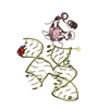
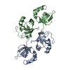
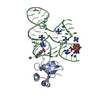
 Citation Citation | Journal: Acta Crystallogr D Biol Crystallogr / Year: 2015 Title: Combination of X-ray crystallography, SAXS and DEER to obtain the structure of the FnIII-3,4 domains of integrin α6β4. Authors: Noelia Alonso-García / Inés García-Rubio / José A Manso / Rubén M Buey / Hector Urien / Arnoud Sonnenberg / Gunnar Jeschke / José M de Pereda /    Abstract: Integrin α6β4 is a major component of hemidesmosomes that mediate the stable anchorage of epithelial cells to the underlying basement membrane. Integrin α6β4 has also been implicated in cell ...Integrin α6β4 is a major component of hemidesmosomes that mediate the stable anchorage of epithelial cells to the underlying basement membrane. Integrin α6β4 has also been implicated in cell proliferation and migration and in carcinoma progression. The third and fourth fibronectin type III domains (FnIII-3,4) of integrin β4 mediate binding to the hemidesmosomal proteins BPAG1e and BPAG2, and participate in signalling. Here, it is demonstrated that X-ray crystallography, small-angle X-ray scattering and double electron-electron resonance (DEER) complement each other to solve the structure of the FnIII-3,4 region. The crystal structures of the individual FnIII-3 and FnIII-4 domains were solved and the relative arrangement of the FnIII domains was elucidated by combining DEER with site-directed spin labelling. Multiple structures of the interdomain linker were modelled by Monte Carlo methods complying with DEER constraints, and the final structures were selected against experimental scattering data. FnIII-3,4 has a compact and cambered flat structure with an evolutionary conserved surface that is likely to correspond to a protein-interaction site. Finally, this hybrid method is of general application for the study of other macromolecules and complexes. |
 Contact author Contact author |
|
- Structure visualization
| Structure viewer | Molecule: MolmilJmol/JSmol |
|---|
- Downloads & links
-Data source
| SASBDB page |  SASDAT6 SASDAT6 |
|---|
-Related structure data


-External links
| Related items in Molecule of the Month |
|---|


-Models
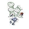
| Model #219 |   Type: dummy / Software: DAMMIF / Radius of dummy atoms: 1.50 A / Chi-square value: 1.976836  Search similar-shape structures of this assembly by Omokage search (details) Search similar-shape structures of this assembly by Omokage search (details) |
|---|---|
| Model #220 |   Type: atomic / Software: CRYSOL / Chi-square value: 1.297321 Search similar-shape structures of this assembly by Omokage search (details) |
-Sample
| Sample | Name: Integrin beta4, fragment of the cytoplasmic region encompassing the third and fourth FnIII domains Specimen concentration: 1.50-48.60 |
|---|---|
| Buffer | Name: Sodium Phosphate / Concentration: 20.00 mM / pH: 7.5 / Composition: 150 mM NaCl, 5% glycerol, 3 mM DTT |
| Entity #133 | Type: protein / Description: Integrin beta-4 / Formula weight: 23.359 / Num. of mol.: 1 / Source: Homo sapiens / References: UniProt: P16144 Sequence: GSHMVPDTPT RLVFSALGPT SLRVSWQEPR CERPLQGYSV EYQLLNGGEL HRLNIPNPAQ TSVVVEDLLP NHSYVFRVRA QSQEGWGRER EGVITIESQV HPQSPLCPLP GSAFTLSTPS APGPLVFTAL SPDSLQLSWE RPRRPNGDIV GYLVTCEMAQ GGGPATAFRV ...Sequence: GSHMVPDTPT RLVFSALGPT SLRVSWQEPR CERPLQGYSV EYQLLNGGEL HRLNIPNPAQ TSVVVEDLLP NHSYVFRVRA QSQEGWGRER EGVITIESQV HPQSPLCPLP GSAFTLSTPS APGPLVFTAL SPDSLQLSWE RPRRPNGDIV GYLVTCEMAQ GGGPATAFRV DGDSPESRLT VPGLSENVPY KFKVQARTTE GFGPEREGII TIES |
-Experimental information
| Beam | Instrument name: PETRA III P12 / City: Hamburg / 国: Germany  / Type of source: X-ray synchrotronSynchrotron / Wavelength: 0.12 Å / Dist. spec. to detc.: 3.1 mm / Type of source: X-ray synchrotronSynchrotron / Wavelength: 0.12 Å / Dist. spec. to detc.: 3.1 mm | |||||||||||||||||||||
|---|---|---|---|---|---|---|---|---|---|---|---|---|---|---|---|---|---|---|---|---|---|---|
| Detector | Name: Pilatus 2M | |||||||||||||||||||||
| Scan | 
| |||||||||||||||||||||
| Distance distribution function P(R) | 
| |||||||||||||||||||||
| Result |  Comments: Combined use of SAXS, crystal structures, site directed spin labeling, double electron-electron resonance (DEER), and molecular modeling to analyze the structure of FnIII-3,4.
|
