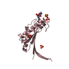
- PDB-4tse: Crystal Structure of the Mib Repeat Domain of Mind bomb 1 -
+
Open data
ID or keywords:
Loading...
-
Basic information
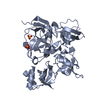
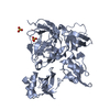
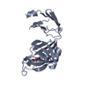
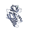
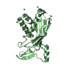
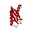
Entry
Database: PDB / ID: 4tse
Title
Crystal Structure of the Mib Repeat Domain of Mind bomb 1
Components
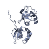
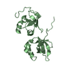
E3 ubiquitin-protein ligase MIB1
Keywords
LIGASE / E3 ubiquitin ligase / Notch pathway
Function / homology
Function and homology information
neural tube formation / blood vessel development / heart looping / positive regulation of endocytosis / centriolar satellite / negative regulation of neuron differentiation / somitogenesis / Notch signaling pathway / Constitutive Signaling by NOTCH1 HD Domain Mutants / NOTCH2 Activation and Transmission of Signal to the Nucleus ...neural tube formation / blood vessel development / heart looping / positive regulation of endocytosis / centriolar satellite / negative regulation of neuron differentiation / somitogenesis / Notch signaling pathway / Constitutive Signaling by NOTCH1 HD Domain Mutants / NOTCH2 Activation and Transmission of Signal to the Nucleus / Activated NOTCH1 Transmits Signal to the Nucleus / NOTCH3 Activation and Transmission of Signal to the Nucleus / RING-type E3 ubiquitin transferase / neuron differentiation / Constitutive Signaling by NOTCH1 PEST Domain Mutants / Constitutive Signaling by NOTCH1 HD+PEST Domain Mutants / endocytosis / ubiquitin-protein transferase activity / ubiquitin protein ligase activity / cytoplasmic vesicle / ubiquitin-dependent protein catabolic process / in utero embryonic development / protein ubiquitination / centrosome / zinc ion binding / plasma membrane / cytosol Similarity search - Function
E3ubiquitin-proteinligaseMIB1 / DAPK-interacting protein 1 / DIP-1 / Mind bomb homolog 1 / Zinc finger ZZ type with ankyrin repeat ...DAPK-interacting protein 1 / DIP-1 / Mind bomb homolog 1 / Zinc finger ZZ type with ankyrin repeat domain protein 2
Mass: 18500.529 Da / Num. of mol.: 2 / Fragment: UNP residues 239-409 Source method: isolated from a genetically manipulated source Source: (gene. exp.) Homo sapiens (human) / Gene: MIB1, DIP1, KIAA1323, ZZANK2 / Plasmid: pTD68 / Production host: Escherichia coli (E. coli) / Strain (production host): BL21(DE3) References: UniProt: Q86YT6, Ligases; Forming carbon-nitrogen bonds; Acid-amino-acid ligases (peptide synthases)
In the structure databanks used in Yorodumi, some data are registered as the other names, "COVID-19 virus" and "2019-nCoV". Here are the details of the virus and the list of structure data.
Jan 31, 2019. EMDB accession codes are about to change! (news from PDBe EMDB page)
EMDB accession codes are about to change! (news from PDBe EMDB page)
The allocation of 4 digits for EMDB accession codes will soon come to an end. Whilst these codes will remain in use, new EMDB accession codes will include an additional digit and will expand incrementally as the available range of codes is exhausted. The current 4-digit format prefixed with “EMD-” (i.e. EMD-XXXX) will advance to a 5-digit format (i.e. EMD-XXXXX), and so on. It is currently estimated that the 4-digit codes will be depleted around Spring 2019, at which point the 5-digit format will come into force.
The EM Navigator/Yorodumi systems omit the EMD- prefix.
Related info.:Q: What is EMD? / ID/Accession-code notation in Yorodumi/EM Navigator
Yorodumi is a browser for structure data from EMDB, PDB, SASBDB, etc.
This page is also the successor to EM Navigator detail page, and also detail information page/front-end page for Omokage search.
The word "yorodu" (or yorozu) is an old Japanese word meaning "ten thousand". "mi" (miru) is to see.
Related info.:EMDB / PDB / SASBDB / Comparison of 3 databanks / Yorodumi Search / Aug 31, 2016. New EM Navigator & Yorodumi / Yorodumi Papers / Jmol/JSmol / Function and homology information / Changes in new EM Navigator and Yorodumi
 Movie
Movie Controller


 Open data
 Basic information
 Components
 Keywords
 LIGASE /
  Function and homology information
 Authors
 United States, 1items
  Citation
 Structure visualization
 Downloads & links
 Other downloads


 PDBj
PDBj


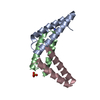
 Assembly
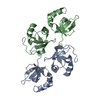

 Mass: 18.015 Da / Num. of mol.: 240 / Source method: isolated from a natural source / Formula: H2O
 Sample preparation
 Processing