 Movie
Movie Controller


+ Open data

- Basic information
| Entry | Database: PDB / ID: 6mog | ||||||
|---|---|---|---|---|---|---|---|
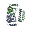
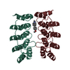
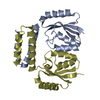
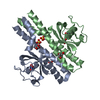
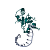
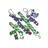
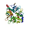
| Title | Dimeric DARPin C_R3 | ||||||
 Components Components | DARPin C_R3 | ||||||
 Keywords Keywords |  BIOSYNTHETIC PROTEIN / DARPin BIOSYNTHETIC PROTEIN / DARPin | ||||||
| Function / homology | Ankyrin repeat-containing domain / Serine Threonine Protein Phosphatase 5, Tetratricopeptide repeat / Alpha Horseshoe / Mainly Alpha / TRIETHYLENE GLYCOL Function and homology information Function and homology information | ||||||
| Biological species | synthetic construct (others) | ||||||
| Method | X-RAY DIFFRACTION / SYNCHROTRON / MOLECULAR REPLACEMENT / Resolution: 1.21 Å | ||||||
 Authors Authors | Jude, K.M. / Mohan, K. / Garcia, K.C. | ||||||
| Funding support |  United States, 1items United States, 1items
| ||||||
 Citation Citation | Journal: Science / Year: 2019 Title: Topological control of cytokine receptor signaling induces differential effects in hematopoiesis. Authors: Mohan, K. / Ueda, G. / Kim, A.R. / Jude, K.M. / Fallas, J.A. / Guo, Y. / Hafer, M. / Miao, Y. / Saxton, R.A. / Piehler, J. / Sankaran, V.G. / Baker, D. / Garcia, K.C. | ||||||
| History |
|
- Structure visualization
| Structure viewer | Molecule: MolmilJmol/JSmol |
|---|
- Downloads & links
-Download
| PDBx/mmCIF format | 6mog.cif.gz | 190.7 KB | Display | PDBx/mmCIF format |
|---|---|---|---|---|
| PDB format | pdb6mog.ent.gz | 156.5 KB | Display | PDB format |
| PDBx/mmJSON format | 6mog.json.gz | Tree view | PDBx/mmJSON format | |
| Others |  Other downloads Other downloads |
-Validation report
| Arichive directory | https://data.pdbj.org/pub/pdb/validation_reports/mo/6mogftp://data.pdbj.org/pub/pdb/validation_reports/mo/6mog | HTTPS FTP |
|---|
-Related structure data
| Related structure data |  6moeC 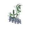 6mofC  6mohC 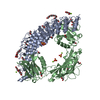 6moiC 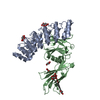 6mojC 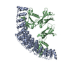 6mokC  6molC C: citing same article ( |
|---|---|
| Similar structure data | |
| Experimental dataset #1 | Data reference: 10.15785/SBGRID/624 / Data set type: diffraction image data |


-Links
 PDBj
PDBj- Assembly
| Deposited unit | 
| ||||||||
|---|---|---|---|---|---|---|---|---|---|
| 1 | 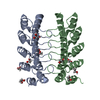
| ||||||||
| Unit cell |
|
-Components
| #1: Protein | Mass: 17565.109 Da / Num. of mol.: 2 Source method: isolated from a genetically manipulated source Source: (gene. exp.) synthetic construct (others) / Production host:  Escherichia coli (E. coli) Escherichia coli (E. coli)#2: Chemical | ChemComp-EDO / Ethylene glycol  Mass: 62.068 Da / Num. of mol.: 10 / Source method: obtained synthetically / Formula: C2H6O2 Mass: 62.068 Da / Num. of mol.: 10 / Source method: obtained synthetically / Formula: C2H6O2#3: Chemical | ChemComp-PGE / | Polyethylene glycol  Mass: 150.173 Da / Num. of mol.: 1 / Source method: obtained synthetically / Formula: C6H14O4 Mass: 150.173 Da / Num. of mol.: 1 / Source method: obtained synthetically / Formula: C6H14O4#4: Water | ChemComp-HOH / | Water Mass: 18.015 Da / Num. of mol.: 180 / Source method: isolated from a natural source / Formula: H2O Mass: 18.015 Da / Num. of mol.: 180 / Source method: isolated from a natural source / Formula: H2O |
|---|
-Experimental details
-Experiment
| Experiment | Method: X-RAY DIFFRACTION / Number of used crystals: 1 |
|---|
- Sample preparation
| Crystal | Density Matthews: 2.03 Å3/Da / Density % sol: 39.36 % |
|---|---|
| Crystal grow | Temperature: 295 K / Method: vapor diffusion, sitting drop / pH: 8.5 Details: 0.1 M Tris pH 8.5, 25% PEG 3350, 1 mM reduced glutathione, 1 mM oxidized glutathione, 25% ethylene glycol cryoprotectant |
-Data collection
| Diffraction | Mean temperature: 100 K / Serial crystal experiment: N |
|---|---|
| Diffraction source | Source: SYNCHROTRON / Site: SSRL / Beamline: BL12-2 / Wavelength: 0.97946 Å |
| Detector | Type: DECTRIS PILATUS 6M / Detector: PIXEL / Date: Jul 23, 2017 |
| Radiation | Protocol: SINGLE WAVELENGTH / Monochromatic (M) / Laue (L): M / Scattering type: x-ray |
| Radiation wavelength | Wavelength: 0.97946 Å / Relative weight: 1 |
| Reflection | Resolution: 1.21→33.25 Å / Num. obs: 83274 / % possible obs: 88.2 % / Redundancy: 6.8 % / Biso Wilson estimate: 14.14 Å2 / CC1/2: 0.999 / Rmerge(I) obs: 0.05076 / Rpim(I) all: 0.0208 / Net I/σ(I): 14.8 |
| Reflection shell | Resolution: 1.21→1.253 Å / Redundancy: 6.7 % / Rmerge(I) obs: 1.564 / Mean I/σ(I) obs: 1.34 / Num. unique obs: 8248 / CC1/2: 0.669 / Rpim(I) all: 0.6433 / % possible all: 72 |
- Processing
| Software |
| |||||||||||||||||||||||||||||||||||||||||||||||||||||||||||||||||||||||||||||||||||||||||||
|---|---|---|---|---|---|---|---|---|---|---|---|---|---|---|---|---|---|---|---|---|---|---|---|---|---|---|---|---|---|---|---|---|---|---|---|---|---|---|---|---|---|---|---|---|---|---|---|---|---|---|---|---|---|---|---|---|---|---|---|---|---|---|---|---|---|---|---|---|---|---|---|---|---|---|---|---|---|---|---|---|---|---|---|---|---|---|---|---|---|---|---|---|
| Refinement | Method to determine structure: MOLECULAR REPLACEMENT Starting model: predicted model Resolution: 1.21→33.248 Å / SU ML: 0.12 / Cross valid method: FREE R-VALUE / σ(F): 0 / Phase error: 21.16
| |||||||||||||||||||||||||||||||||||||||||||||||||||||||||||||||||||||||||||||||||||||||||||
| Solvent computation | Shrinkage radii: 0.9 Å / VDW probe radii: 1.11 Å | |||||||||||||||||||||||||||||||||||||||||||||||||||||||||||||||||||||||||||||||||||||||||||
| Refinement step | Cycle: LAST / Resolution: 1.21→33.248 Å
| |||||||||||||||||||||||||||||||||||||||||||||||||||||||||||||||||||||||||||||||||||||||||||
| Refine LS restraints |
| |||||||||||||||||||||||||||||||||||||||||||||||||||||||||||||||||||||||||||||||||||||||||||
| LS refinement shell |
|
