 Movie
Movie Controller

[English] 日本語
 Yorodumi
- PDB-3ub9: Periplasmic portion of the Helicobacter pylori chemoreceptor TlpB... -
+ Open data

- Basic information
| Entry | Database: PDB / ID: 3ub9 | ||||||
|---|---|---|---|---|---|---|---|
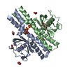
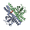
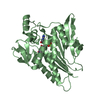
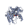
| Title | Periplasmic portion of the Helicobacter pylori chemoreceptor TlpB with hydroxyurea bound | ||||||
 Components Components | chemoreceptor TlpB | ||||||
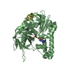
 Keywords Keywords |  MEMBRANE PROTEIN / homodimer / four-helix bundle / PAS domain MEMBRANE PROTEIN / homodimer / four-helix bundle / PAS domain | ||||||
| Function / homology |  Function and homology information Function and homology information | ||||||
| Biological species |   Helicobacter pylori (bacteria) Helicobacter pylori (bacteria) | ||||||
| Method | X-RAY DIFFRACTION / SYNCHROTRON / MOLECULAR REPLACEMENT / Resolution: 1.42 Å | ||||||
 Authors Authors | Henderson, J.N. / Sweeney, E.G. / Goers, J. / Wreden, C. / Hicks, K.G. / Parthasarathy, R. / Guillemin, K.J. / Remington, S.J. | ||||||
 Citation Citation | Journal: Structure / Year: 2012 Title: Structure and proposed mechanism for the pH-sensing Helicobacter pylori chemoreceptor TlpB. Authors: Goers Sweeney, E. / Henderson, J.N. / Goers, J. / Wreden, C. / Hicks, K.G. / Foster, J.K. / Parthasarathy, R. / Remington, S.J. / Guillemin, K. | ||||||
| History |
|
- Structure visualization
| Structure viewer | Molecule: MolmilJmol/JSmol |
|---|
- Downloads & links
-Download
| PDBx/mmCIF format | 3ub9.cif.gz | 167.2 KB | Display | PDBx/mmCIF format |
|---|---|---|---|---|
| PDB format | pdb3ub9.ent.gz | 133 KB | Display | PDB format |
| PDBx/mmJSON format | 3ub9.json.gz | Tree view | PDBx/mmJSON format | |
| Others |  Other downloads Other downloads |
-Validation report
| Arichive directory | https://data.pdbj.org/pub/pdb/validation_reports/ub/3ub9ftp://data.pdbj.org/pub/pdb/validation_reports/ub/3ub9 | HTTPS FTP |
|---|
-Related structure data


-Links
 PDBj
PDBj

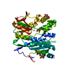
- Assembly
| Deposited unit | 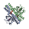
| ||||||||
|---|---|---|---|---|---|---|---|---|---|
| 1 |
| ||||||||
| Unit cell |
| ||||||||
| Details | biological unit is the same as asym. |
-Components
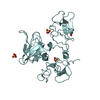
| #1: Protein | Mass: 20687.516 Da / Num. of mol.: 2 / Fragment: Periplasmic portion Source method: isolated from a genetically manipulated source Source: (gene. exp.) Helicobacter pylori (bacteria) / Strain: SS1 / Plasmid: pBH4 / Production host: Escherichia coli (E. coli) / Strain (production host): BL21(DE3) / References: UniProt: B6JPK4*PLUS#2: Chemical | Hydroxycarbamide  Mass: 76.055 Da / Num. of mol.: 2 / Source method: obtained synthetically / Formula: CH4N2O2 / Comment: medication*YM Mass: 76.055 Da / Num. of mol.: 2 / Source method: obtained synthetically / Formula: CH4N2O2 / Comment: medication*YM#3: Chemical | Sulfate  Mass: 96.063 Da / Num. of mol.: 3 / Source method: obtained synthetically / Formula: SO4 Mass: 96.063 Da / Num. of mol.: 3 / Source method: obtained synthetically / Formula: SO4#4: Chemical | ChemComp-GOL / Glycerol  Mass: 92.094 Da / Num. of mol.: 5 / Source method: obtained synthetically / Formula: C3H8O3 Mass: 92.094 Da / Num. of mol.: 5 / Source method: obtained synthetically / Formula: C3H8O3#5: Water | ChemComp-HOH / | Water Mass: 18.015 Da / Num. of mol.: 473 / Source method: isolated from a natural source / Formula: H2O Mass: 18.015 Da / Num. of mol.: 473 / Source method: isolated from a natural source / Formula: H2O |
|---|
-Experimental details
-Experiment
| Experiment | Method: X-RAY DIFFRACTION / Number of used crystals: 1 |
|---|
- Sample preparation
| Crystal | Density Matthews: 3.71 Å3/Da / Density % sol: 66.83 % |
|---|---|
| Crystal grow | Temperature: 277 K / Method: vapor diffusion, hanging drop / pH: 6.5 Details: 0.1 M Bis-Tris pH 7.0, 2.0 M ammonium sulfate, vapor diffusion, hanging drop, temperature 277K |
-Data collection
| Diffraction | Mean temperature: 100 K | ||||||||||||||||||||||||||||||||||||||||||||||||||||||||||||||||||||||||||||||||||||||||||||||||||||||||||||||||
|---|---|---|---|---|---|---|---|---|---|---|---|---|---|---|---|---|---|---|---|---|---|---|---|---|---|---|---|---|---|---|---|---|---|---|---|---|---|---|---|---|---|---|---|---|---|---|---|---|---|---|---|---|---|---|---|---|---|---|---|---|---|---|---|---|---|---|---|---|---|---|---|---|---|---|---|---|---|---|---|---|---|---|---|---|---|---|---|---|---|---|---|---|---|---|---|---|---|---|---|---|---|---|---|---|---|---|---|---|---|---|---|---|---|
| Diffraction source | Source: SYNCHROTRON / Site: ALS  / Beamline: 5.0.2 / Wavelength: 0.97 Å / Beamline: 5.0.2 / Wavelength: 0.97 Å | ||||||||||||||||||||||||||||||||||||||||||||||||||||||||||||||||||||||||||||||||||||||||||||||||||||||||||||||||
| Detector | Type: ADSC QUANTUM 315 / Detector: CCD / Date: Nov 15, 2008 / Details: Rh/Pt coated Si | ||||||||||||||||||||||||||||||||||||||||||||||||||||||||||||||||||||||||||||||||||||||||||||||||||||||||||||||||
| Radiation | Monochromator: DOUBLE-CRYSTAL, SI(111) / Protocol: SINGLE WAVELENGTH / Monochromatic (M) / Laue (L): M / Scattering type: x-ray | ||||||||||||||||||||||||||||||||||||||||||||||||||||||||||||||||||||||||||||||||||||||||||||||||||||||||||||||||
| Radiation wavelength | Wavelength: 0.97 Å / Relative weight: 1 | ||||||||||||||||||||||||||||||||||||||||||||||||||||||||||||||||||||||||||||||||||||||||||||||||||||||||||||||||
| Reflection | Resolution: 1.42→50 Å / Num. all: 116572 / Num. obs: 113775 / % possible obs: 97.6 % / Observed criterion σ(I): -3 / Redundancy: 3.5 % / Rmerge(I) obs: 0.062 / Χ2: 0.916 / Net I/σ(I): 13.1 | ||||||||||||||||||||||||||||||||||||||||||||||||||||||||||||||||||||||||||||||||||||||||||||||||||||||||||||||||
| Reflection shell |
|
- Processing
| Software |
| ||||||||||||||||||||||||||||||||||||||||||||||||||||||||||||||||||||||||||||||||||||||||||||||||||||
|---|---|---|---|---|---|---|---|---|---|---|---|---|---|---|---|---|---|---|---|---|---|---|---|---|---|---|---|---|---|---|---|---|---|---|---|---|---|---|---|---|---|---|---|---|---|---|---|---|---|---|---|---|---|---|---|---|---|---|---|---|---|---|---|---|---|---|---|---|---|---|---|---|---|---|---|---|---|---|---|---|---|---|---|---|---|---|---|---|---|---|---|---|---|---|---|---|---|---|---|---|---|
| Refinement | Method to determine structure: MOLECULAR REPLACEMENT / Resolution: 1.42→40.65 Å / Cor.coef. Fo:Fc: 0.972 / Cor.coef. Fo:Fc free: 0.965 / WRfactor Rfree: 0.1539 / WRfactor Rwork: 0.1289 / Occupancy max: 1 / Occupancy min: 0.3 / FOM work R set: 0.9376 / SU B: 1.039 / SU ML: 0.019 / SU R Cruickshank DPI: 0.0388 / SU Rfree: 0.0393 / Cross valid method: THROUGHOUT / σ(F): 0 / ESU R Free: 0.039 / Stereochemistry target values: MAXIMUM LIKELIHOOD Details: HYDROGENS HAVE BEEN ADDED IN THE RIDING POSITIONS U VALUES : REFINED INDIVIDUALLY
| ||||||||||||||||||||||||||||||||||||||||||||||||||||||||||||||||||||||||||||||||||||||||||||||||||||
| Solvent computation | Ion probe radii: 0.8 Å / Shrinkage radii: 0.8 Å / VDW probe radii: 1.4 Å / Solvent model: MASK | ||||||||||||||||||||||||||||||||||||||||||||||||||||||||||||||||||||||||||||||||||||||||||||||||||||
| Displacement parameters | Biso max: 61.11 Å2 / Biso mean: 17.1805 Å2 / Biso min: 5.98 Å2
| ||||||||||||||||||||||||||||||||||||||||||||||||||||||||||||||||||||||||||||||||||||||||||||||||||||
| Refinement step | Cycle: LAST / Resolution: 1.42→40.65 Å
| ||||||||||||||||||||||||||||||||||||||||||||||||||||||||||||||||||||||||||||||||||||||||||||||||||||
| Refine LS restraints |
| ||||||||||||||||||||||||||||||||||||||||||||||||||||||||||||||||||||||||||||||||||||||||||||||||||||
| LS refinement shell | Resolution: 1.421→1.458 Å / Total num. of bins used: 20
|
