Movie
Movie Controller

[English] 日本語
 Yorodumi
- PDB-9avl: Structure of human calcium-sensing receptor in complex with Gi3 p... -
+ Open data

- Basic information
| Entry | Database: PDB / ID: 9avl | ||||||
|---|---|---|---|---|---|---|---|
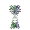
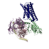
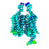
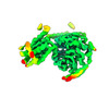
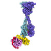
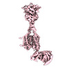
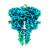
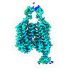
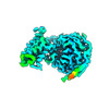
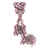
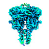
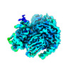
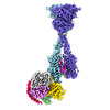
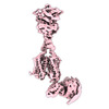
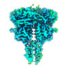
| Title | Structure of human calcium-sensing receptor in complex with Gi3 protein in nanodiscs | ||||||
 Components Components |
| ||||||
 Keywords Keywords |  MEMBRANE PROTEIN / Calcium-sensing receptor / G-protein-coupled receptor / G protein / signal transduction MEMBRANE PROTEIN / Calcium-sensing receptor / G-protein-coupled receptor / G protein / signal transduction | ||||||
| Function / homology |  Function and homology information Function and homology informationbile acid secretion / chemosensory behavior / cellular response to peptide / response to fibroblast growth factor / cellular response to vitamin D / phosphatidylinositol phospholipase C activity / negative regulation of adenylate cyclase activity / Class C/3 (Metabotropic glutamate/pheromone receptors) / regulation of potassium ion transmembrane transport / calcium ion import ...bile acid secretion / chemosensory behavior / cellular response to peptide / response to fibroblast growth factor / cellular response to vitamin D / phosphatidylinositol phospholipase C activity / negative regulation of adenylate cyclase activity / Class C/3 (Metabotropic glutamate/pheromone receptors) / regulation of potassium ion transmembrane transport / calcium ion import / GTP metabolic process / positive regulation of positive chemotaxis / fat pad development / dopamine receptor signaling pathway / amino acid binding / cellular response to hepatocyte growth factor stimulus / branching morphogenesis of an epithelial tube / positive regulation of calcium ion import / regulation of calcium ion transport / positive regulation of macroautophagy / cellular response to low-density lipoprotein particle stimulus / detection of calcium ion / Adenylate cyclase inhibitory pathway / anatomical structure morphogenesis / axon terminus / JNK cascade / positive regulation of vasoconstriction / chloride transmembrane transport / adenylate cyclase-inhibiting G protein-coupled receptor signaling pathway / ossification / G protein-coupled receptor activity / G protein-coupled receptor binding / response to ischemia / cellular response to glucose stimulus / G-protein beta/gamma-subunit complex binding / adenylate cyclase-modulating G protein-coupled receptor signaling pathway / G beta:gamma signalling through PLC beta / Presynaptic function of Kainate receptors / Thromboxane signalling through TP receptor / G-protein activation / Activation of G protein gated Potassium channels / Inhibition of voltage gated Ca2+ channels via Gbeta/gamma subunits / positive regulation of insulin secretion / Prostacyclin signalling through prostacyclin receptor / Glucagon signaling in metabolic regulation / G beta:gamma signalling through CDC42 / ADP signalling through P2Y purinoceptor 12 / G beta:gamma signalling through BTK / Adrenaline,noradrenaline inhibits insulin secretion / Glucagon-type ligand receptors / Vasopressin regulates renal water homeostasis via Aquaporins / G alpha (z) signalling events / cellular response to catecholamine stimulus / Glucagon-like Peptide-1 (GLP1) regulates insulin secretion / intracellular calcium ion homeostasis / ADORA2B mediated anti-inflammatory cytokines production / ADP signalling through P2Y purinoceptor 1 / adenylate cyclase-activating dopamine receptor signaling pathway / G beta:gamma signalling through PI3Kgamma / cellular response to prostaglandin E stimulus / Cooperation of PDCL (PhLP1) and TRiC/CCT in G-protein beta folding / vasodilation / GPER1 signaling / G-protein beta-subunit binding / GDP binding / heterotrimeric G-protein complex / G alpha (12/13) signalling events / signaling receptor complex adaptor activity / integrin binding / Thrombin signalling through proteinase activated receptors (PARs) / GTPase binding / phospholipase C-activating G protein-coupled receptor signaling pathway / Ca2+ pathway / midbody / G alpha (i) signalling events / fibroblast proliferation / cellular response to hypoxia / G alpha (s) signalling events / G alpha (q) signalling events / basolateral plasma membrane / vesicle / transmembrane transporter binding / Extra-nuclear estrogen signaling / positive regulation of ERK1 and ERK2 cascade / apical plasma membrane / G protein-coupled receptor signaling pathway / cell cycle / lysosomal membrane / cell division / focal adhesion / GTPase activity / centrosome / neuronal cell body / calcium ion binding / positive regulation of cell population proliferation / protein-containing complex binding / positive regulation of gene expression / nucleolus / GTP binding / protein kinase bindingSimilarity search - Function | ||||||
| Biological species |  Homo sapiens (human) Homo sapiens (human) | ||||||
| Method | ELECTRON MICROSCOPY / single particle reconstruction / cryo EM / Resolution: 3.8 Å | ||||||
 Authors Authors | Zuo, H. / Park, J. / Frangaj, A. / Ye, J. / Lu, G. / Manning, J.J. / Asher, W.B. / Lu, Z. / Hu, G. / Wang, L. ...Zuo, H. / Park, J. / Frangaj, A. / Ye, J. / Lu, G. / Manning, J.J. / Asher, W.B. / Lu, Z. / Hu, G. / Wang, L. / Mendez, J. / Eng, E. / Zhang, Z. / Lin, X. / Grasucci, R. / Hendrickson, W.A. / Clarke, O.B. / Javitch, J.A. / Conigrave, A.D. / Fan, Q.R. | ||||||
| Funding support |  United States, 1items United States, 1items
| ||||||
 Citation Citation | Journal: Nature / Year: 2024 Title: Promiscuous G-protein activation by the calcium-sensing receptor. Authors: Hao Zuo / Jinseo Park / Aurel Frangaj / Jianxiang Ye / Guanqi Lu / Jamie J Manning / Wesley B Asher / Zhengyuan Lu / Guo-Bin Hu / Liguo Wang / Joshua Mendez / Edward Eng / Zhening Zhang / ...Authors: Hao Zuo / Jinseo Park / Aurel Frangaj / Jianxiang Ye / Guanqi Lu / Jamie J Manning / Wesley B Asher / Zhengyuan Lu / Guo-Bin Hu / Liguo Wang / Joshua Mendez / Edward Eng / Zhening Zhang / Xin Lin / Robert Grassucci / Wayne A Hendrickson / Oliver B Clarke / Jonathan A Javitch / Arthur D Conigrave / Qing R Fan /  Abstract: The human calcium-sensing receptor (CaSR) detects fluctuations in the extracellular Ca concentration and maintains Ca homeostasis. It also mediates diverse cellular processes not associated with Ca ...The human calcium-sensing receptor (CaSR) detects fluctuations in the extracellular Ca concentration and maintains Ca homeostasis. It also mediates diverse cellular processes not associated with Ca balance. The functional pleiotropy of CaSR arises in part from its ability to signal through several G-protein subtypes. We determined structures of CaSR in complex with G proteins from three different subfamilies: G, G and G. We found that the homodimeric CaSR of each complex couples to a single G protein through a common mode. This involves the C-terminal helix of each Gα subunit binding to a shallow pocket that is formed in one CaSR subunit by all three intracellular loops (ICL1-ICL3), an extended transmembrane helix 3 and an ordered C-terminal region. G-protein binding expands the transmembrane dimer interface, which is further stabilized by phospholipid. The restraint imposed by the receptor dimer, in combination with ICL2, enables G-protein activation by facilitating conformational transition of Gα. We identified a single Gα residue that determines G and G versus G selectivity. The length and flexibility of ICL2 allows CaSR to bind all three Gα subtypes, thereby conferring capacity for promiscuous G-protein coupling. | ||||||
| History |
|
- Structure visualization
| Structure viewer | Molecule: MolmilJmol/JSmol |
|---|
- Downloads & links
-Download
| PDBx/mmCIF format | 9avl.cif.gz | 424.1 KB | Display | PDBx/mmCIF format |
|---|---|---|---|---|
| PDB format | pdb9avl.ent.gz | Display | PDB format | |
| PDBx/mmJSON format | 9avl.json.gz | Tree view | PDBx/mmJSON format | |
| Others |  Other downloads Other downloads |
-Validation report
| Arichive directory | https://data.pdbj.org/pub/pdb/validation_reports/av/9avlftp://data.pdbj.org/pub/pdb/validation_reports/av/9avl | HTTPS FTP |
|---|
-Related structure data
| Related structure data | 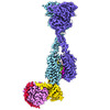 43908MC  9asbC  9avgC 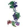 9axfC 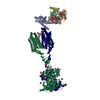 9ayfC C: citing same article ( M: map data used to model this data |
|---|---|
| Similar structure data |


-Links
 PDBj
PDBj


- Assembly
| Deposited unit | 
|
|---|---|
| 1 |
|
-Components
-Protein , 1 types, 2 molecules QR
| #1: Protein | Mass: 102864.617 Da / Num. of mol.: 2 Source method: isolated from a genetically manipulated source Details: The CaSR construct consists of residues 1-903 and a Flag tag inserted after the signal peptide. Source: (gene. exp.) Homo sapiens (human) / Gene: CASR, GPRC2A, PCAR1 / Cell line (production host): HEK293 GnTI- / Production host: Homo sapiens (human) / References: UniProt: P41180 |
|---|
-Guanine nucleotide-binding protein ... , 3 types, 3 molecules ABG
| #2: Protein | Mass: 40584.156 Da / Num. of mol.: 1 / Mutation: S47N, G203A, E245A, A326S Source method: isolated from a genetically manipulated source Details: G(i3)alpha contains four dominant negative mutations, S47N, G203A, E245A, and A326S Source: (gene. exp.) Homo sapiens (human) / Gene: GNAI3 / Cell line (production host): HEK293 GnTI- / Production host: Homo sapiens (human) / References: UniProt: P08754 |
|---|---|
| #3: Protein | Mass: 38367.969 Da / Num. of mol.: 1 Source method: isolated from a genetically manipulated source Details: GNB2 has N-terminal Flag tag inserted after the initial Met. Source: (gene. exp.) Homo sapiens (human) / Gene: GNB2 / Cell line (production host): HEK293 GnTI- / Production host: Homo sapiens (human) / References: UniProt: P62879 |
| #4: Protein | Mass: 7861.143 Da / Num. of mol.: 1 Source method: isolated from a genetically manipulated source Source: (gene. exp.) Homo sapiens (human) / Gene: GNG2 / Cell line (production host): HEK293 GnTI- / Production host: Homo sapiens (human) / References: UniProt: P59768 |
-Sugars , 2 types, 8 molecules 
| #5: Polysaccharide | 2-acetamido-2-deoxy-beta-D-glucopyranose-(1-4)-2-acetamido-2-deoxy-beta-D-glucopyranose / Mass: 424.401 Da / Num. of mol.: 4Source method: isolated from a genetically manipulated source #6: Sugar | ChemComp-NAG / N-Acetylglucosamine Type: D-saccharide, beta linking / Mass: 221.208 Da / Num. of mol.: 4 / Source method: obtained synthetically / Formula: C8H15NO6 Type: D-saccharide, beta linking / Mass: 221.208 Da / Num. of mol.: 4 / Source method: obtained synthetically / Formula: C8H15NO6 |
|---|
-Non-polymers , 6 types, 14 molecules 


| #7: Chemical |  Type: L-peptide linking / Mass: 216.236 Da / Num. of mol.: 2 / Source method: obtained synthetically / Formula: C12H12N2O2 Type: L-peptide linking / Mass: 216.236 Da / Num. of mol.: 2 / Source method: obtained synthetically / Formula: C12H12N2O2#8: Chemical | Phosphate Mass: 94.971 Da / Num. of mol.: 2 / Source method: obtained synthetically / Formula: PO4 Mass: 94.971 Da / Num. of mol.: 2 / Source method: obtained synthetically / Formula: PO4#9: Chemical | ChemComp-CA /  Mass: 40.078 Da / Num. of mol.: 6 / Source method: obtained synthetically / Formula: Ca Mass: 40.078 Da / Num. of mol.: 6 / Source method: obtained synthetically / Formula: Ca#10: Chemical |  Mass: 303.826 Da / Num. of mol.: 2 / Source method: obtained synthetically / Formula: C18H22ClNO Mass: 303.826 Da / Num. of mol.: 2 / Source method: obtained synthetically / Formula: C18H22ClNO#11: Chemical | ChemComp-Y01 / |  Mass: 486.726 Da / Num. of mol.: 1 / Source method: obtained synthetically / Formula: C31H50O4 Mass: 486.726 Da / Num. of mol.: 1 / Source method: obtained synthetically / Formula: C31H50O4#12: Chemical | ChemComp-A1AF7 / ( | Mass: 749.007 Da / Num. of mol.: 1 / Source method: obtained synthetically / Formula: C40H77O10P |
|---|
-Details
| Has ligand of interest | N |
|---|
-Experimental details
-Experiment
| Experiment | Method: ELECTRON MICROSCOPY |
|---|---|
| EM experiment | Aggregation state: PARTICLE / 3D reconstruction method: single particle reconstruction |
- Sample preparation
| Component | Name: Human CaSR in complex with Gi3 protein / Type: COMPLEX / Entity ID: #1-#4 / Source: RECOMBINANT | |||||||||||||||||||||||||||||||||||
|---|---|---|---|---|---|---|---|---|---|---|---|---|---|---|---|---|---|---|---|---|---|---|---|---|---|---|---|---|---|---|---|---|---|---|---|---|
| Molecular weight | Value: 0.288 MDa / Experimental value: NO | |||||||||||||||||||||||||||||||||||
| Source (natural) | Organism: Homo sapiens (human) | |||||||||||||||||||||||||||||||||||
| Source (recombinant) | Organism: Homo sapiens (human) / Cell: HEK293 GnTI- | |||||||||||||||||||||||||||||||||||
| Buffer solution | pH: 7.5 | |||||||||||||||||||||||||||||||||||
| Buffer component |
| |||||||||||||||||||||||||||||||||||
| Specimen | Conc.: 3.5 mg/ml / Embedding applied: NO / Shadowing applied: NO / Staining applied: NO / Vitrification applied: YES | |||||||||||||||||||||||||||||||||||
| Specimen support | Grid material: GOLD / Grid mesh size: 300 divisions/in. / Grid type: Quantifoil R0.6/1 | |||||||||||||||||||||||||||||||||||
| Vitrification | Instrument: FEI VITROBOT MARK IV / Cryogen name: ETHANE / Humidity: 100 % / Chamber temperature: 277 K Details: The sample was blotted for 6s before plunge-frozen. |
- Electron microscopy imaging
| Experimental equipment |  Model: Titan Krios / Image courtesy: FEI Company |
|---|---|
| Microscopy | Model: FEI TITAN KRIOS |
| Electron gun | Electron source: FIELD EMISSION GUN / Accelerating voltage: 300 kV / Illumination mode: FLOOD BEAM |
| Electron lens | Mode: BRIGHT FIELDBright-field microscopy / Nominal magnification: 105000 X / Nominal defocus max: 1800 nm / Nominal defocus min: 1000 nm / Cs: 2.7 mm / C2 aperture diameter: 100 µm / Alignment procedure: COMA FREE |
| Specimen holder | Cryogen: NITROGEN / Specimen holder model: FEI TITAN KRIOS AUTOGRID HOLDER / Temperature (max): 100 K |
| Image recording | Average exposure time: 2.5 sec. / Electron dose: 70.14 e/Å2 / Film or detector model: GATAN K3 BIOQUANTUM (6k x 4k) / Num. of grids imaged: 2 / Num. of real images: 16504 |
| EM imaging optics | Energyfilter name: GIF Bioquantum / Energyfilter slit width: 20 eV |
| Image scans | Width: 5760 / Height: 4092 |
- Processing
| EM software |
| ||||||||||||||||||||||||||||||||||||
|---|---|---|---|---|---|---|---|---|---|---|---|---|---|---|---|---|---|---|---|---|---|---|---|---|---|---|---|---|---|---|---|---|---|---|---|---|---|
| CTF correction | Type: PHASE FLIPPING AND AMPLITUDE CORRECTION | ||||||||||||||||||||||||||||||||||||
| Particle selection | Num. of particles selected: 2792317 | ||||||||||||||||||||||||||||||||||||
| Symmetry | Point symmetry: C1 (asymmetric) | ||||||||||||||||||||||||||||||||||||
| 3D reconstruction | Resolution: 3.8 Å / Resolution method: FSC 0.143 CUT-OFF / Num. of particles: 55985 / Symmetry type: POINT | ||||||||||||||||||||||||||||||||||||
| Atomic model building | Protocol: FLEXIBLE FIT / Space: REAL | ||||||||||||||||||||||||||||||||||||
| Atomic model building | 3D fitting-ID: 1 / Source name: PDB / Type: experimental model
| ||||||||||||||||||||||||||||||||||||
| Refine LS restraints |
|