 Movie
Movie Controller


+ Open data

- Basic information
| Entry | Database: PDB / ID: 8uue | |||||||||||||||
|---|---|---|---|---|---|---|---|---|---|---|---|---|---|---|---|---|
| Title | Glycine-bound GluN1a-3A LBD heterotetramer (local refinement) | |||||||||||||||
 Components Components |
| |||||||||||||||
 Keywords Keywords |  MEMBRANE PROTEIN / Channel / receptor MEMBRANE PROTEIN / Channel / receptor | |||||||||||||||
| Function / homology |  Function and homology information Function and homology informationnegative regulation of dendritic spine development / excitatory chemical synaptic transmission / Synaptic adhesion-like molecules / propylene metabolic process / response to glycine / glutamate-gated calcium ion channel activity / Assembly and cell surface presentation of NMDA receptors / regulation of monoatomic cation transmembrane transport / glutamate receptor activity / Neurexins and neuroligins ...negative regulation of dendritic spine development / excitatory chemical synaptic transmission / Synaptic adhesion-like molecules / propylene metabolic process / response to glycine / glutamate-gated calcium ion channel activity / Assembly and cell surface presentation of NMDA receptors / regulation of monoatomic cation transmembrane transport / glutamate receptor activity / Neurexins and neuroligins / NMDA glutamate receptor activity / NMDA selective glutamate receptor complex / calcium ion transmembrane import into cytosol / protein heterotetramerization / glutamate binding / positive regulation of reactive oxygen species biosynthetic process / glycine binding / positive regulation of calcium ion transport into cytosol / regulation of neuronal synaptic plasticity / Negative regulation of NMDA receptor-mediated neuronal transmission / dendrite development / Unblocking of NMDA receptors, glutamate binding and activation / monoatomic cation transmembrane transport / positive regulation of excitatory postsynaptic potential / ligand-gated monoatomic ion channel activity / Long-term potentiation / monoatomic cation transport / excitatory synapse / calcium ion homeostasis / synaptic cleft / prepulse inhibition / EPHB-mediated forward signaling / excitatory postsynaptic potential / presynaptic modulation of chemical synaptic transmission / ionotropic glutamate receptor signaling pathway / Ras activation upon Ca2+ influx through NMDA receptor / regulation of membrane potential / positive regulation of synaptic transmission, glutamatergic / transmitter-gated monoatomic ion channel activity involved in regulation of postsynaptic membrane potential / protein phosphatase 2A binding / synaptic membrane / synaptic transmission, glutamatergic / postsynaptic density membrane / brain development / regulation of synaptic plasticity / modulation of chemical synaptic transmission / calcium channel activity / visual learning / terminal bouton / rhythmic process / calcium ion transport / synaptic vesicle / presynapse / signaling receptor activity / amyloid-beta binding / chemical synaptic transmission / RAF/MAP kinase cascade / postsynaptic membrane / response to ethanol / dendritic spine / postsynaptic density / calmodulin binding / neuron projection / dendrite / neuronal cell body / glutamatergic synapse / synapse / calcium ion binding / protein-containing complex binding / endoplasmic reticulum membrane / cell surface / positive regulation of transcription by RNA polymerase II / membrane / identical protein binding / plasma membrane / cytoplasmSimilarity search - Function | |||||||||||||||
| Biological species |  Homo sapiens (human) Homo sapiens (human) | |||||||||||||||
| Method | ELECTRON MICROSCOPY / single particle reconstruction / cryo EM / Resolution: 3.96 Å | |||||||||||||||
 Authors Authors | Michalski, K. / Furukawa, H. | |||||||||||||||
| Funding support |  United States, 4items United States, 4items
| |||||||||||||||
 Citation Citation | Journal: Sci Adv / Year: 2024 Title: Structure and function of GluN1-3A NMDA receptor excitatory glycine receptor channel. Authors: Kevin Michalski / Hiro Furukawa / Abstract: -methyl-d-aspartate receptors (NMDARs) and other ionotropic glutamate receptors (iGluRs) mediate most of the excitatory signaling in the mammalian brains in response to the neurotransmitter glutamate. ...-methyl-d-aspartate receptors (NMDARs) and other ionotropic glutamate receptors (iGluRs) mediate most of the excitatory signaling in the mammalian brains in response to the neurotransmitter glutamate. Uniquely, NMDARs composed of GluN1 and GluN3 are activated exclusively by glycine, the neurotransmitter conventionally mediating inhibitory signaling when it binds to pentameric glycine receptors. The GluN1-3 NMDARs are vital for regulating neuronal excitability, circuit function, and specific behaviors, yet our understanding of their functional mechanism at the molecular level has remained limited. Here, we present cryo-electron microscopy structures of GluN1-3A NMDARs bound to an antagonist, CNQX, and an agonist, glycine. The structures show a 1-3-1-3 subunit heterotetrameric arrangement and an unprecedented pattern of GluN3A subunit orientation shift between the glycine-bound and CNQX-bound structures. Site-directed disruption of the unique subunit interface in the glycine-bound structure mitigated desensitization. Our study provides a foundation for understanding the distinct structural dynamics of GluN3 that are linked to the unique function of GluN1-3 NMDARs. | |||||||||||||||
| History |
|
- Structure visualization
| Structure viewer | Molecule: MolmilJmol/JSmol |
|---|
- Downloads & links
-Download
| PDBx/mmCIF format | 8uue.cif.gz | 216 KB | Display | PDBx/mmCIF format |
|---|---|---|---|---|
| PDB format | pdb8uue.ent.gz | 168.9 KB | Display | PDB format |
| PDBx/mmJSON format | 8uue.json.gz | Tree view | PDBx/mmJSON format | |
| Others |  Other downloads Other downloads |
-Validation report
| Arichive directory | https://data.pdbj.org/pub/pdb/validation_reports/uu/8uueftp://data.pdbj.org/pub/pdb/validation_reports/uu/8uue | HTTPS FTP |
|---|
-Related structure data
| Related structure data |  42580MC 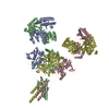 8uswC 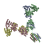 8usxC M: map data used to model this data C: citing same article ( |
|---|---|
| Similar structure data |


-Links
 PDBj
PDBj


- Assembly
| Deposited unit | 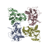
|
|---|---|
| 1 |
|
-Components
| #1: Protein | Mass: 45902.477 Da / Num. of mol.: 2 Source method: isolated from a genetically manipulated source Source: (gene. exp.) Homo sapiens (human) / Gene: GRIN1, NMDAR1 / Production host:   Spodoptera frugiperda (fall armyworm) / References: UniProt: Q05586 Spodoptera frugiperda (fall armyworm) / References: UniProt: Q05586#2: Protein | Mass: 45535.320 Da / Num. of mol.: 2 Source method: isolated from a genetically manipulated source Source: (gene. exp.) Homo sapiens (human) / Gene: GRIN3A, KIAA1973 / Production host: Spodoptera frugiperda (fall armyworm) / References: UniProt: Q8TCU5 |
|---|
-Experimental details
-Experiment
| Experiment | Method: ELECTRON MICROSCOPY |
|---|---|
| EM experiment | Aggregation state: PARTICLE / 3D reconstruction method: single particle reconstruction |
- Sample preparation
| Component | Name: Glycine-bound GluN1-3A LBD heterotetramer (local refinement) Type: COMPLEX / Entity ID: all / Source: RECOMBINANT |
|---|---|
| Molecular weight | Value: 0.4 MDa / Experimental value: NO |
| Source (natural) | Organism: Homo sapiens (human) |
| Source (recombinant) | Organism: Spodoptera frugiperda (fall armyworm) |
| Buffer solution | pH: 7.5 |
| Specimen | Embedding applied: NO / Shadowing applied: NO / Staining applied: NO / Vitrification applied: YES |
| Vitrification | Cryogen name: ETHANE |
- Electron microscopy imaging
| Experimental equipment |  Model: Titan Krios / Image courtesy: FEI Company |
|---|---|
| Microscopy | Model: FEI TITAN KRIOS |
| Electron gun | Electron source: FIELD EMISSION GUN / Accelerating voltage: 300 kV / Illumination mode: OTHER |
| Electron lens | Mode: OTHER / Nominal defocus max: 2200 nm / Nominal defocus min: 800 nm |
| Image recording | Electron dose: 60 e/Å2 / Film or detector model: GATAN K3 (6k x 4k) |
- Processing
| CTF correction | Type: NONE |
|---|---|
| 3D reconstruction | Resolution: 3.96 Å / Resolution method: FSC 0.143 CUT-OFF / Num. of particles: 225900 / Symmetry type: POINT |
