Protein or peptide: Glutamate receptor ionotropic, NMDA 1
Protein or peptide: Glutamate receptor ionotropic, NMDA 3A
Keywords
Channel / receptor / MEMBRANE PROTEIN
Function / homology
Function and homology information
negative regulation of dendritic spine development / excitatory chemical synaptic transmission / Synaptic adhesion-like molecules / propylene metabolic process / response to glycine / glutamate-gated calcium ion channel activity / Assembly and cell surface presentation of NMDA receptors / regulation of monoatomic cation transmembrane transport / glutamate receptor activity / Neurexins and neuroligins ...negative regulation of dendritic spine development / excitatory chemical synaptic transmission / Synaptic adhesion-like molecules / propylene metabolic process / response to glycine / glutamate-gated calcium ion channel activity / Assembly and cell surface presentation of NMDA receptors / regulation of monoatomic cation transmembrane transport / glutamate receptor activity / Neurexins and neuroligins / NMDA glutamate receptor activity / NMDA selective glutamate receptor complex / calcium ion transmembrane import into cytosol / protein heterotetramerization / glutamate binding / positive regulation of reactive oxygen species biosynthetic process / glycine binding / positive regulation of calcium ion transport into cytosol / regulation of neuronal synaptic plasticity / Negative regulation of NMDA receptor-mediated neuronal transmission / dendrite development / Unblocking of NMDA receptors, glutamate binding and activation / monoatomic cation transmembrane transport / positive regulation of excitatory postsynaptic potential / ligand-gated monoatomic ion channel activity / Long-term potentiation / monoatomic cation transport / excitatory synapse / calcium ion homeostasis / synaptic cleft / prepulse inhibition / EPHB-mediated forward signaling / excitatory postsynaptic potential / presynaptic modulation of chemical synaptic transmission / ionotropic glutamate receptor signaling pathway / Ras activation upon Ca2+ influx through NMDA receptor / regulation of membrane potential / positive regulation of synaptic transmission, glutamatergic / transmitter-gated monoatomic ion channel activity involved in regulation of postsynaptic membrane potential / protein phosphatase 2A binding / synaptic membrane / synaptic transmission, glutamatergic / postsynaptic density membrane / brain development / regulation of synaptic plasticity / modulation of chemical synaptic transmission / calcium channel activity / visual learning / terminal bouton / rhythmic process / calcium ion transport / synaptic vesicle / presynapse / signaling receptor activity / amyloid-beta binding / chemical synaptic transmission / RAF/MAP kinase cascade / postsynaptic membrane / response to ethanol / dendritic spine / postsynaptic density / calmodulin binding / neuron projection / dendrite / neuronal cell body / glutamatergic synapse / synapse / calcium ion binding / protein-containing complex binding / endoplasmic reticulum membrane / cell surface / positive regulation of transcription by RNA polymerase II / membrane / identical protein binding / plasma membrane / cytoplasm Similarity search - Function
Bacterial extracellular solute-binding proteins, family 3 / Solute-binding protein family 3/N-terminal domain of MltF / Ionotropic glutamate receptor, metazoa / Ligated ion channel L-glutamate- and glycine-binding site / : / Ligand-gated ion channel / Ionotropic glutamate receptor, L-glutamate and glycine-binding domain / Ligated ion channel L-glutamate- and glycine-binding site / Ionotropic glutamate receptor / Eukaryotic homologues of bacterial periplasmic substrate binding proteins. ...Bacterial extracellular solute-binding proteins, family 3 / Solute-binding protein family 3/N-terminal domain of MltF / Ionotropic glutamate receptor, metazoa / Ligated ion channel L-glutamate- and glycine-binding site / : / Ligand-gated ion channel / Ionotropic glutamate receptor, L-glutamate and glycine-binding domain / Ligated ion channel L-glutamate- and glycine-binding site / Ionotropic glutamate receptor / Eukaryotic homologues of bacterial periplasmic substrate binding proteins. / Receptor, ligand binding region / Receptor family ligand binding region / Periplasmic binding protein-like I Similarity search - Domain/homology
National Institutes of Health/National Institute of Mental Health (NIH/NIMH)
NS11745
United States
National Institutes of Health/National Institute of Mental Health (NIH/NIMH)
MH085926
United States
National Institutes of Health/National Institute of Mental Health (NIH/NIMH)
F32MH121061
United States
National Institutes of Health/National Institute of Mental Health (NIH/NIMH)
NS113632
United States
Citation
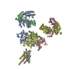
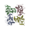
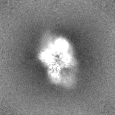
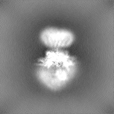
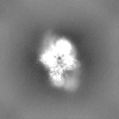
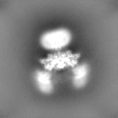
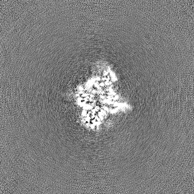
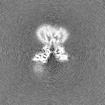
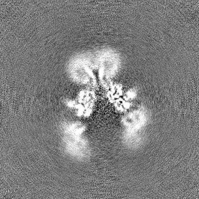
Journal: Sci Adv / Year: 2024 Title: Structure and function of GluN1-3A NMDA receptor excitatory glycine receptor channel. Authors: Kevin Michalski / Hiro Furukawa / Abstract: -methyl-d-aspartate receptors (NMDARs) and other ionotropic glutamate receptors (iGluRs) mediate most of the excitatory signaling in the mammalian brains in response to the neurotransmitter glutamate. ...-methyl-d-aspartate receptors (NMDARs) and other ionotropic glutamate receptors (iGluRs) mediate most of the excitatory signaling in the mammalian brains in response to the neurotransmitter glutamate. Uniquely, NMDARs composed of GluN1 and GluN3 are activated exclusively by glycine, the neurotransmitter conventionally mediating inhibitory signaling when it binds to pentameric glycine receptors. The GluN1-3 NMDARs are vital for regulating neuronal excitability, circuit function, and specific behaviors, yet our understanding of their functional mechanism at the molecular level has remained limited. Here, we present cryo-electron microscopy structures of GluN1-3A NMDARs bound to an antagonist, CNQX, and an agonist, glycine. The structures show a 1-3-1-3 subunit heterotetrameric arrangement and an unprecedented pattern of GluN3A subunit orientation shift between the glycine-bound and CNQX-bound structures. Site-directed disruption of the unique subunit interface in the glycine-bound structure mitigated desensitization. Our study provides a foundation for understanding the distinct structural dynamics of GluN3 that are linked to the unique function of GluN1-3 NMDARs.
In the structure databanks used in Yorodumi, some data are registered as the other names, "COVID-19 virus" and "2019-nCoV". Here are the details of the virus and the list of structure data.
Jan 31, 2019. EMDB accession codes are about to change! (news from PDBe EMDB page)
EMDB accession codes are about to change! (news from PDBe EMDB page)
The allocation of 4 digits for EMDB accession codes will soon come to an end. Whilst these codes will remain in use, new EMDB accession codes will include an additional digit and will expand incrementally as the available range of codes is exhausted. The current 4-digit format prefixed with “EMD-” (i.e. EMD-XXXX) will advance to a 5-digit format (i.e. EMD-XXXXX), and so on. It is currently estimated that the 4-digit codes will be depleted around Spring 2019, at which point the 5-digit format will come into force.
The EM Navigator/Yorodumi systems omit the EMD- prefix.
Related info.:Q: What is EMD? / ID/Accession-code notation in Yorodumi/EM Navigator
Yorodumi is a browser for structure data from EMDB, PDB, SASBDB, etc.
This page is also the successor to EM Navigator detail page, and also detail information page/front-end page for Omokage search.
The word "yorodu" (or yorozu) is an old Japanese word meaning "ten thousand". "mi" (miru) is to see.
Related info.:EMDB / PDB / SASBDB / Comparison of 3 databanks / Yorodumi Search / Aug 31, 2016. New EM Navigator & Yorodumi / Yorodumi Papers / Jmol/JSmol / Function and homology information / Changes in new EM Navigator and Yorodumi
 Movie
Movie Controller


 Open data
 Basic information
 Map data
 Sample
 Keywords
 receptor /
  Function and homology information
 Authors
 United States, 4 items
  Citation
 Structure visualization
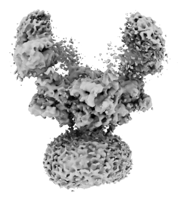
 Downloads & links
 emd_42522.png
 http://ftp.pdbj.org/pub/emdb/structures/EMD-42522
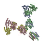


 Z (Sec.)
 Y (Row.)
 X (Col.)


 Sample components

 Processing
 Electron microscopy
