Movie
Movie Controller


+ Open data

- Basic information
| Entry | Database: PDB / ID: 8u3z | ||||||
|---|---|---|---|---|---|---|---|
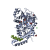
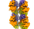
| Title | Model of TTLL6 bound to microtubule from composite map | ||||||
 Components Components |
| ||||||
 Keywords Keywords |  LIGASE / tubulin / post-translational modifications / microtubules / Tubulin Tyrosine Ligase Like family enzymes LIGASE / tubulin / post-translational modifications / microtubules / Tubulin Tyrosine Ligase Like family enzymes | ||||||
| Function / homology |  Function and homology information Function and homology informationpositive regulation of cilium movement / protein-glutamic acid ligase activity / tubulin-glutamic acid ligase activity / Carboxyterminal post-translational modifications of tubulin / protein polyglutamylation / regulation of cilium beat frequency involved in ciliary motility / 9+0 non-motile cilium / microtubule severing / Ligases; Forming carbon-nitrogen bonds; Acid-amino-acid ligases (peptide synthases) / odontoblast differentiation ...positive regulation of cilium movement / protein-glutamic acid ligase activity / tubulin-glutamic acid ligase activity / Carboxyterminal post-translational modifications of tubulin / protein polyglutamylation / regulation of cilium beat frequency involved in ciliary motility / 9+0 non-motile cilium / microtubule severing / Ligases; Forming carbon-nitrogen bonds; Acid-amino-acid ligases (peptide synthases) / odontoblast differentiation / Post-chaperonin tubulin folding pathway / Carboxyterminal post-translational modifications of tubulin / Microtubule-dependent trafficking of connexons from Golgi to the plasma membrane / Cilium Assembly / Sealing of the nuclear envelope (NE) by ESCRT-III / Intraflagellar transport / cytoskeleton-dependent intracellular transport / Formation of tubulin folding intermediates by CCT/TriC / Gap junction assembly / COPI-independent Golgi-to-ER retrograde traffic / natural killer cell mediated cytotoxicity / microtubule bundle formation / Assembly and cell surface presentation of NMDA receptors / Kinesins / GTPase activating protein binding / COPI-dependent Golgi-to-ER retrograde traffic / intercellular bridge / regulation of synapse organization / nuclear envelope lumen / cytoplasmic microtubule / Recycling pathway of L1 / MHC class I protein binding / RHOH GTPase cycle / RHO GTPases activate IQGAPs / spindle assembly / cellular response to interleukin-4 / microtubule-based process / Hedgehog 'off' state / COPI-mediated anterograde transport / Activation of AMPK downstream of NMDARs / Mitotic Prometaphase / EML4 and NUDC in mitotic spindle formation / Loss of Nlp from mitotic centrosomes / Loss of proteins required for interphase microtubule organization from the centrosome / Recruitment of mitotic centrosome proteins and complexes / Resolution of Sister Chromatid Cohesion / Recruitment of NuMA to mitotic centrosomes / Anchoring of the basal body to the plasma membrane / HSP90 chaperone cycle for steroid hormone receptors (SHR) in the presence of ligand / MHC class II antigen presentation / tubulin binding / AURKA Activation by TPX2 / ciliary basal body / Translocation of SLC2A4 (GLUT4) to the plasma membrane / RHO GTPases Activate Formins / Hydrolases; Acting on acid anhydrides; Acting on GTP to facilitate cellular and subcellular movement / PKR-mediated signaling / cilium / mitotic spindle / structural constituent of cytoskeleton / microtubule cytoskeleton organization / Aggrephagy / cytoplasmic ribonucleoprotein granule / HCMV Early Events / Separation of Sister Chromatids / The role of GTSE1 in G2/M progression after G2 checkpoint / microtubule cytoskeleton / Regulation of PLK1 Activity at G2/M Transition / azurophil granule lumen / double-stranded RNA binding / mitotic cell cycle / cell body / microtubule / Potential therapeutics for SARS / cytoskeleton / membrane raft / protein domain specific binding / cell division / GTPase activity / ubiquitin protein ligase binding / Neutrophil degranulation / protein-containing complex binding / GTP binding / structural molecule activity / protein-containing complex / extracellular exosome / extracellular region / ATP binding / metal ion binding / nucleus / cytosol / cytoplasmSimilarity search - Function | ||||||
| Biological species |  Mus musculus (house mouse) Mus musculus (house mouse) Homo sapiens (human) Homo sapiens (human) | ||||||
| Method | ELECTRON MICROSCOPY / single particle reconstruction / cryo EM / Resolution: 3.6 Å | ||||||
 Authors Authors | Mahalingan, K.K. / Grotjahn, D. / Li, Y. / Lander, G.C. / Zehr, E.A. / Roll-Mecak, A. | ||||||
| Funding support |  United States, 1items United States, 1items
| ||||||
 Citation Citation | Journal: Nat Chem Biol / Year: 2024 Title: Structural basis for α-tubulin-specific and modification state-dependent glutamylation. Authors: Kishore K Mahalingan / Danielle A Grotjahn / Yan Li / Gabriel C Lander / Elena A Zehr / Antonina Roll-Mecak / Abstract: Microtubules have spatiotemporally complex posttranslational modification patterns. Tubulin tyrosine ligase-like (TTLL) enzymes introduce the most prevalent modifications on α-tubulin and β-tubulin. ...Microtubules have spatiotemporally complex posttranslational modification patterns. Tubulin tyrosine ligase-like (TTLL) enzymes introduce the most prevalent modifications on α-tubulin and β-tubulin. How TTLLs specialize for specific substrate recognition and ultimately modification-pattern generation is largely unknown. TTLL6, a glutamylase implicated in ciliopathies, preferentially modifies tubulin α-tails in microtubules. Cryo-electron microscopy, kinetic analysis and single-molecule biochemistry reveal an unprecedented quadrivalent recognition that ensures simultaneous readout of microtubule geometry and posttranslational modification status. By binding to a β-tubulin subunit, TTLL6 modifies the α-tail of the longitudinally adjacent tubulin dimer. Spanning two tubulin dimers along and across protofilaments (PFs) ensures fidelity of recognition of both the α-tail and the microtubule. Moreover, TTLL6 reads out and is stimulated by glutamylation of the β-tail of the laterally adjacent tubulin dimer, mediating crosstalk between α-tail and β-tail. This positive feedback loop can generate localized microtubule glutamylation patterns. Our work uncovers general principles that generate tubulin chemical and topographic complexity. | ||||||
| History |
|
- Structure visualization
| Structure viewer | Molecule: MolmilJmol/JSmol |
|---|
- Downloads & links
-Download
| PDBx/mmCIF format | 8u3z.cif.gz | 425.3 KB | Display | PDBx/mmCIF format |
|---|---|---|---|---|
| PDB format | pdb8u3z.ent.gz | 338.1 KB | Display | PDB format |
| PDBx/mmJSON format | 8u3z.json.gz | Tree view | PDBx/mmJSON format | |
| Others |  Other downloads Other downloads |
-Validation report
| Arichive directory | https://data.pdbj.org/pub/pdb/validation_reports/u3/8u3zftp://data.pdbj.org/pub/pdb/validation_reports/u3/8u3z | HTTPS FTP |
|---|
-Related structure data
| Related structure data |  41090MC  8t42C C: citing same article ( M: map data used to model this data |
|---|---|
| Similar structure data | |
| Experimental dataset #1 | Data reference: 10.6019/EMPIAR-11798 / Data set type: EMPIAR Details: Cryo-EM micrographs of human alpha1B/BetaI+BetaIVb microtubules bound to GMPCPP decorated with TTLL6 Metadata reference: 10.6019/EMPIAR-11798 |


-Links
 PDBj
PDBj


- Assembly
| Deposited unit | 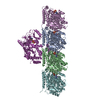
|
|---|---|
| 1 |
|
-Components
-Protein , 3 types, 5 molecules AFBEN
| #1: Protein | Mass: 50204.445 Da / Num. of mol.: 2 / Source method: isolated from a natural source / Source: (natural) Homo sapiens (human) / Cell line: tsA201 / Organ: kidney / Tissue: kidney / References: UniProt: P68363#2: Protein | Mass: 49717.629 Da / Num. of mol.: 2 / Source method: isolated from a natural source / Source: (natural) Homo sapiens (human) / Cell line: tsA201 / Organ: kidney / Tissue: kidney / References: UniProt: P07437#3: Protein | | Mass: 94601.188 Da / Num. of mol.: 1 Source method: isolated from a genetically manipulated source Source: (gene. exp.) Mus musculus (house mouse) / Gene: Ttll6 / Plasmid: P7XNH3 / Production host:  Escherichia coli (E. coli) / Strain (production host): Arctic Express DE3 Escherichia coli (E. coli) / Strain (production host): Arctic Express DE3References: UniProt: A4Q9E8, Ligases; Forming carbon-nitrogen bonds; Acid-amino-acid ligases (peptide synthases) |
|---|
-Non-polymers , 4 types, 10 molecules 


| #4: Chemical | Guanosine triphosphate Mass: 523.180 Da / Num. of mol.: 2 / Source method: obtained synthetically / Formula: C10H16N5O14P3 / Comment: GTP, energy-carrying molecule*YM Mass: 523.180 Da / Num. of mol.: 2 / Source method: obtained synthetically / Formula: C10H16N5O14P3 / Comment: GTP, energy-carrying molecule*YM#5: Chemical | ChemComp-MG /  Mass: 24.305 Da / Num. of mol.: 5 / Source method: obtained synthetically / Formula: Mg Mass: 24.305 Da / Num. of mol.: 5 / Source method: obtained synthetically / Formula: Mg#6: Chemical |  Mass: 521.208 Da / Num. of mol.: 2 / Source method: obtained synthetically / Formula: C11H18N5O13P3 / Feature type: SUBJECT OF INVESTIGATION / Comment: GMP-CPP, energy-carrying molecule analogue*YM Mass: 521.208 Da / Num. of mol.: 2 / Source method: obtained synthetically / Formula: C11H18N5O13P3 / Feature type: SUBJECT OF INVESTIGATION / Comment: GMP-CPP, energy-carrying molecule analogue*YM#7: Chemical | ChemComp-ATP / | Adenosine triphosphate Mass: 507.181 Da / Num. of mol.: 1 / Source method: obtained synthetically / Formula: C10H16N5O13P3 / Comment: ATP, energy-carrying molecule*YM Mass: 507.181 Da / Num. of mol.: 1 / Source method: obtained synthetically / Formula: C10H16N5O13P3 / Comment: ATP, energy-carrying molecule*YM |
|---|
-Details
| Has ligand of interest | Y |
|---|
-Experimental details
-Experiment
| Experiment | Method: ELECTRON MICROSCOPY |
|---|---|
| EM experiment | Aggregation state: FILAMENT / 3D reconstruction method: single particle reconstruction |
- Sample preparation
| Component |
| |||||||||||||||||||||||||||||||||||
|---|---|---|---|---|---|---|---|---|---|---|---|---|---|---|---|---|---|---|---|---|---|---|---|---|---|---|---|---|---|---|---|---|---|---|---|---|
| Molecular weight |
| |||||||||||||||||||||||||||||||||||
| Source (natural) |
| |||||||||||||||||||||||||||||||||||
| Source (recombinant) | Organism: Escherichia coli (E. coli) / Strain: Arctic Express DE3 / Plasmid: P7XNH3 | |||||||||||||||||||||||||||||||||||
| Buffer solution | pH: 7 | |||||||||||||||||||||||||||||||||||
| Buffer component |
| |||||||||||||||||||||||||||||||||||
| Specimen | Conc.: 0.1 mg/ml / Embedding applied: NO / Shadowing applied: NO / Staining applied: NO / Vitrification applied: YES / Details: TTLL6 decorated microtubules heterogeneously | |||||||||||||||||||||||||||||||||||
| Specimen support | Grid material: GOLD / Grid mesh size: 300 divisions/in. / Grid type: Quantifoil R1.2/1.3 | |||||||||||||||||||||||||||||||||||
| Vitrification | Instrument: FEI VITROBOT MARK IV / Cryogen name: ETHANE / Humidity: 100 % / Chamber temperature: 303 K |
- Electron microscopy imaging
| Experimental equipment |  Model: Talos Arctica / Image courtesy: FEI Company |
|---|---|
| Microscopy | Model: FEI TALOS ARCTICA |
| Electron gun | Electron source: FIELD EMISSION GUN / Accelerating voltage: 200 kV / Illumination mode: FLOOD BEAM |
| Electron lens | Mode: BRIGHT FIELDBright-field microscopy / Nominal magnification: 36000 X / Nominal defocus max: 2000 nm / Nominal defocus min: 1000 nm / Cs: 2.7 mm / Alignment procedure: COMA FREE |
| Image recording | Average exposure time: 9.75 sec. / Electron dose: 42 e/Å2 / Detector mode: COUNTING / Film or detector model: GATAN K2 SUMMIT (4k x 4k) |
| Image scans | Movie frames/image: 39 / Used frames/image: 1-39 |
- Processing
| EM software |
| ||||||||||||||||||||||||||||
|---|---|---|---|---|---|---|---|---|---|---|---|---|---|---|---|---|---|---|---|---|---|---|---|---|---|---|---|---|---|
| CTF correction | Type: PHASE FLIPPING AND AMPLITUDE CORRECTION | ||||||||||||||||||||||||||||
| Particle selection | Num. of particles selected: 59055 Details: Due to highly heterogeneous TTLL decoration microtubules were manually selected. 59044 overlapping segments with a box size of 568 pixels were extracted every 82 A | ||||||||||||||||||||||||||||
| Symmetry | Point symmetry: C1 (asymmetric) | ||||||||||||||||||||||||||||
| 3D reconstruction | Resolution: 3.6 Å / Resolution method: FSC 0.143 CUT-OFF / Num. of particles: 392289 / Algorithm: FOURIER SPACE / Num. of class averages: 1 / Symmetry type: POINT | ||||||||||||||||||||||||||||
| Atomic model building | Protocol: FLEXIBLE FIT / Space: REAL / Target criteria: cross-correlation | ||||||||||||||||||||||||||||
| Atomic model building | 3D fitting-ID: 1 / Source name: PDB / Type: experimental model
|