 Movie
Movie Controller


+ Open data

- Basic information
| Entry |  | |||||||||
|---|---|---|---|---|---|---|---|---|---|---|
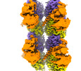
| Title | Map of Tubulin Tyrosine Ligase Like 6 | |||||||||
 Map data Map data | Map of TTLL6 bound to unmodified human microtubules, obtained by focused classification and refinement | |||||||||
 Sample Sample |
| |||||||||
 Keywords Keywords | Tubulin polyglutamylase / tubulin post-translational modifications /  microtubules / TTLL / LIGASE microtubules / TTLL / LIGASE | |||||||||
| Function / homology |  Function and homology information Function and homology informationpositive regulation of cilium movement / protein-glutamic acid ligase activity / tubulin-glutamic acid ligase activity / Carboxyterminal post-translational modifications of tubulin / protein polyglutamylation / regulation of cilium beat frequency involved in ciliary motility / 9+0 non-motile cilium / microtubule severing / Ligases; Forming carbon-nitrogen bonds; Acid-amino-acid ligases (peptide synthases) / microtubule bundle formation ...positive regulation of cilium movement / protein-glutamic acid ligase activity / tubulin-glutamic acid ligase activity / Carboxyterminal post-translational modifications of tubulin / protein polyglutamylation / regulation of cilium beat frequency involved in ciliary motility / 9+0 non-motile cilium / microtubule severing / Ligases; Forming carbon-nitrogen bonds; Acid-amino-acid ligases (peptide synthases) / microtubule bundle formation / tubulin binding / ciliary basal body / cilium / microtubule cytoskeleton organization / microtubule / ATP binding / metal ion binding / cytoplasmSimilarity search - Function | |||||||||
| Biological species |  Mus musculus (house mouse) Mus musculus (house mouse) | |||||||||
| Method | single particle reconstruction / cryo EM / Resolution: 7.2 Å | |||||||||
 Authors Authors | Mahalingan KK / Grotjahn D / Li Y / Lander GC / Zehr EA / Roll-Mecak A | |||||||||
| Funding support |  United States, 1 items United States, 1 items
| |||||||||
 Citation Citation | Journal: Nat Chem Biol / Year: 2024 Title: Structural basis for α-tubulin-specific and modification state-dependent glutamylation. Authors: Kishore K Mahalingan / Danielle A Grotjahn / Yan Li / Gabriel C Lander / Elena A Zehr / Antonina Roll-Mecak / Abstract: Microtubules have spatiotemporally complex posttranslational modification patterns. Tubulin tyrosine ligase-like (TTLL) enzymes introduce the most prevalent modifications on α-tubulin and β-tubulin. ...Microtubules have spatiotemporally complex posttranslational modification patterns. Tubulin tyrosine ligase-like (TTLL) enzymes introduce the most prevalent modifications on α-tubulin and β-tubulin. How TTLLs specialize for specific substrate recognition and ultimately modification-pattern generation is largely unknown. TTLL6, a glutamylase implicated in ciliopathies, preferentially modifies tubulin α-tails in microtubules. Cryo-electron microscopy, kinetic analysis and single-molecule biochemistry reveal an unprecedented quadrivalent recognition that ensures simultaneous readout of microtubule geometry and posttranslational modification status. By binding to a β-tubulin subunit, TTLL6 modifies the α-tail of the longitudinally adjacent tubulin dimer. Spanning two tubulin dimers along and across protofilaments (PFs) ensures fidelity of recognition of both the α-tail and the microtubule. Moreover, TTLL6 reads out and is stimulated by glutamylation of the β-tail of the laterally adjacent tubulin dimer, mediating crosstalk between α-tail and β-tail. This positive feedback loop can generate localized microtubule glutamylation patterns. Our work uncovers general principles that generate tubulin chemical and topographic complexity. | |||||||||
| History |
|
- Structure visualization
| Supplemental images |
|---|

- Downloads & links
-EMDB archive
| Map data | emd_41022.map.gz | 4.4 MB | EMDB map data format | |
|---|---|---|---|---|
| Header (meta data) | emd-41022-v30.xmlemd-41022.xml | 20.1 KB 20.1 KB | Display Display | EMDB header |
| FSC (resolution estimation) | emd_41022_fsc.xmlemd_41022_fsc_2.xml | 20.2 KB 20 KB | Display Display | FSC data file |
| Images |  emd_41022.png emd_41022.png | 44.6 KB | ||
| Masks | emd_41022_msk_1.map | 699 MB | Mask map | |
| Filedesc metadata | emd-41022.cif.gz | 6.3 KB | ||
| Others | emd_41022_half_map_1.map.gzemd_41022_half_map_2.map.gz | 557.9 MB 557.5 MB | ||
| Archive directory |  http://ftp.pdbj.org/pub/emdb/structures/EMD-41022ftp://ftp.pdbj.org/pub/emdb/structures/EMD-41022 http://ftp.pdbj.org/pub/emdb/structures/EMD-41022ftp://ftp.pdbj.org/pub/emdb/structures/EMD-41022 | HTTPS FTP |
-Related structure data


-Links
| EMDB pages | EMDB (EBI/PDBe) / EMDataResource |
|---|
-Map
| File | Download / File: emd_41022.map.gz / Format: CCP4 / Size: 699 MB / Type: IMAGE STORED AS FLOATING POINT NUMBER (4 BYTES) | ||||||||||||||||||||
|---|---|---|---|---|---|---|---|---|---|---|---|---|---|---|---|---|---|---|---|---|---|
| Annotation | Map of TTLL6 bound to unmodified human microtubules, obtained by focused classification and refinement | ||||||||||||||||||||
| Voxel size | X=Y=Z: 1.15 Å | ||||||||||||||||||||
| Density |
| ||||||||||||||||||||
| Symmetry | Space group: 1 | ||||||||||||||||||||
| Details | EMDB XML:
|
-Supplemental data
-Mask #1
| File | emd_41022_msk_1.map | ||||||||||||
|---|---|---|---|---|---|---|---|---|---|---|---|---|---|
| Projections & Slices |
| ||||||||||||
| Density Histograms |
 Z
Z Y
Y X
X


-Half map: First half-map of TTLL6 bound to unmodified human...
| File | emd_41022_half_map_1.map | ||||||||||||
|---|---|---|---|---|---|---|---|---|---|---|---|---|---|
| Annotation | First half-map of TTLL6 bound to unmodified human microtubules, obtained by focused classification and refinement | ||||||||||||
| Projections & Slices |
| ||||||||||||
| Density Histograms |


-Half map: Second half-map of TTLL6 bound to unmodified human...
| File | emd_41022_half_map_2.map | ||||||||||||
|---|---|---|---|---|---|---|---|---|---|---|---|---|---|
| Annotation | Second half-map of TTLL6 bound to unmodified human microtubules, obtained by focused classification and refinement | ||||||||||||
| Projections & Slices |
| ||||||||||||
| Density Histograms |


- Sample components
-Entire : TTLL6 bound to unmodified human microtubules
| Entire | Name: TTLL6 bound to unmodified human microtubules |
|---|---|
| Components |
|
-Supramolecule #1: TTLL6 bound to unmodified human microtubules
| Supramolecule | Name: TTLL6 bound to unmodified human microtubules / type: complex / ID: 1 / Parent: 0 / Macromolecule list: all Details: Microtubules were decorated with TTLL6 on electron microscopy grids |
|---|---|
| Source (natural) | Organism: Mus musculus (house mouse) / Location in cell: cytoplasm |
| Molecular weight | Theoretical: 20 KDa |
-Macromolecule #1: Tubulin Tyrosine Ligase Like 6
| Macromolecule | Name: Tubulin Tyrosine Ligase Like 6 / type: protein_or_peptide / ID: 1 / Enantiomer: LEVO / EC number: ec: 6.3.2.61 |
|---|---|
| Source (natural) | Organism: Mus musculus (house mouse) |
| Recombinant expression | Organism:  |
| Sequence | String: GKKKRKKKRL VINLSNCRYD SVRRAAQQYG LREAGDNDDW TLYWTDYSVS LERVMEMKSY QKINHFPGMS EICRKDLLAR NMSRMLKLFP KDFHFFPRTW CLPADWGDLQ TYSRTRKNKT YICKPDSGCQ GRGIFITRSV KEIKPGEDMI CQLYISKPFI IDGFKFDLRV ...String: GKKKRKKKRL VINLSNCRYD SVRRAAQQYG LREAGDNDDW TLYWTDYSVS LERVMEMKSY QKINHFPGMS EICRKDLLAR NMSRMLKLFP KDFHFFPRTW CLPADWGDLQ TYSRTRKNKT YICKPDSGCQ GRGIFITRSV KEIKPGEDMI CQLYISKPFI IDGFKFDLRV YVLVTSCDPL RVFVYNEGLA RFATTSYSHP NLDNLDEICM HLTNYSINKH SSNFVQDAFS GSKRKLSTFN SYMKTHGYDV EQIWRGIEDV IIKTLISAHP VIKHNYHTCF PSHTLNSACF EILGFDILLD RKLKPWLLEV NHSPSFSTDS KLDKEVKDSL LYDALVLINL GNCDKKKVLE EERQRGRFLQ QCPNREIRLE EVKGFQAMRL QKTEEYEKKN CGGFRLIYPG LNLEKYDKFF QDNSSLFQNT VASRARELYA RQLIQELRQK QEKKVFLKKA RKA UniProtKB: Tubulin polyglutamylase TTLL6 |
-Experimental details
-Structure determination
| Method | cryo EM |
|---|---|
 Processing Processing | single particle reconstruction |
| Aggregation state | particle |
-Sample preparation
| Concentration | 0.1 mg/mL | ||||||||||||||||||
|---|---|---|---|---|---|---|---|---|---|---|---|---|---|---|---|---|---|---|---|
| Buffer | pH: 7 Component:
| ||||||||||||||||||
| Grid | Model: Quantifoil R1.2/1.3 / Material: GOLD / Mesh: 300 / Support film - Material: GOLD / Support film - topology: HOLEY / Pretreatment - Type: GLOW DISCHARGE / Pretreatment - Time: 30 sec. / Pretreatment - Atmosphere: AIR | ||||||||||||||||||
| Vitrification | Cryogen name: ETHANE / Chamber humidity: 100 % / Chamber temperature: 303 K / Instrument: FEI VITROBOT MARK IV Details: The sample was blotted with Whatman #5 blotting paper on both sides of the grid for 3 s with a blot offset of -1 using a Vitrobot Mark IV (Thermo Fisher Scientific) with a chamber set to 30C ...Details: The sample was blotted with Whatman #5 blotting paper on both sides of the grid for 3 s with a blot offset of -1 using a Vitrobot Mark IV (Thermo Fisher Scientific) with a chamber set to 30C at 100% humidity and subsequently plunged into liquid ethane.. | ||||||||||||||||||
| Details | TTLL6 decoration of microtubule was heterogenous |
- Electron microscopy
| Microscope | FEI TALOS ARCTICA |
|---|---|
| Electron beam | Acceleration voltage: 200 kV / Electron source: FIELD EMISSION GUN |
| Electron optics | Illumination mode: FLOOD BEAM / Imaging mode: BRIGHT FIELDBright-field microscopy / Cs: 2.7 mm / Nominal defocus max: 2.0 µm / Nominal defocus min: 1.0 µm / Nominal magnification: 36000 |
| Image recording | Film or detector model: GATAN K2 SUMMIT (4k x 4k) / Detector mode: COUNTING / Digitization - Frames/image: 1-39 / Number real images: 2771 / Average exposure time: 9.75 sec. / Average electron dose: 42.0 e/Å2 |
| Experimental equipment |  Model: Talos Arctica / Image courtesy: FEI Company |
-Image processing
| Particle selection | Number selected: 59044 Details: Due to highly heterogeneous TTLL6 decoration of microtubules microtubules were manually selected. 59044 overlapping segments with a box size of 568 pixels were extracted every 82 A |
|---|---|
| Startup model | Type of model: OTHER Details: Map of TTLL6 bound to unmodified human microtubules |
| Initial angle assignment | Type: MAXIMUM LIKELIHOOD / Software - Name: RELION (ver. 3.0.8) |
| Final 3D classification | Number classes: 2 / Avg.num./class: 196000 / Software - Name: RELION (ver. 3.0.8) |
| Final angle assignment | Type: MAXIMUM LIKELIHOOD / Software - Name: RELION (ver. 3.0.8) |
| Final reconstruction | Number classes used: 1 / Applied symmetry - Point group: C1 (asymmetric) / Algorithm: FOURIER SPACE / Resolution.type: BY AUTHOR / Resolution: 7.2 Å / Resolution method: FSC 0.143 CUT-OFF / Software - Name: RELION (ver. 3.0.8) Details: Map obtained through focused classification with subtraction of microtubule signal, followed by 3D refinement. Number images used: 151716 |
| FSC plot (resolution estimation) |  |
