 Movie
Movie Controller

[English] 日本語
 Yorodumi
- PDB-8k5c: Cryo-EM structure of Acipimox bound human hydroxy-carboxylic acid... -
+ Open data

- Basic information
| Entry | Database: PDB / ID: 8k5c | ||||||
|---|---|---|---|---|---|---|---|
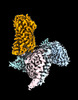
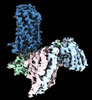
| Title | Cryo-EM structure of Acipimox bound human hydroxy-carboxylic acid receptor 2 (Local refinement) | ||||||
 Components Components | Human hydroxycarboxylic acid receptor 2 | ||||||
 Keywords Keywords |  MEMBRANE PROTEIN / GPCR / G-Protein / acipimox / signaling MEMBRANE PROTEIN / GPCR / G-Protein / acipimox / signaling | ||||||
| Function / homology |  Function and homology informationnicotinic acid receptor activity / Hydroxycarboxylic acid-binding receptors / neutrophil apoptotic process / Class A/1 (Rhodopsin-like receptors) / positive regulation of neutrophil apoptotic process / positive regulation of adiponectin secretion / negative regulation of lipid catabolic process / cell junction / G alpha (i) signalling events / G protein-coupled receptor signaling pathway / plasma membrane Function and homology informationnicotinic acid receptor activity / Hydroxycarboxylic acid-binding receptors / neutrophil apoptotic process / Class A/1 (Rhodopsin-like receptors) / positive regulation of neutrophil apoptotic process / positive regulation of adiponectin secretion / negative regulation of lipid catabolic process / cell junction / G alpha (i) signalling events / G protein-coupled receptor signaling pathway / plasma membraneSimilarity search - Function | ||||||
| Biological species |  Homo sapiens (human) Homo sapiens (human) | ||||||
| Method | ELECTRON MICROSCOPY / single particle reconstruction / cryo EM / Resolution: 3.13 Å | ||||||
 Authors Authors | Park, J.H. / Ishimoto, N. / Park, S.Y. | ||||||
| Funding support | 1items
| ||||||
 Citation Citation | Journal: Nat Commun / Year: 2023 Title: Structural basis for ligand recognition and signaling of hydroxy-carboxylic acid receptor 2. Authors: Jae-Hyun Park / Kouki Kawakami / Naito Ishimoto / Tatsuya Ikuta / Mio Ohki / Toru Ekimoto / Mitsunori Ikeguchi / Dong-Sun Lee / Young-Ho Lee / Jeremy R H Tame / Asuka Inoue / Sam-Yong Park /   Abstract: Hydroxycarboxylic acid receptors (HCAR1, HCAR2, and HCAR3) transduce G signaling upon biding to molecules such as lactic acid, butyric acid and 3-hydroxyoctanoic acid, which are associated with ...Hydroxycarboxylic acid receptors (HCAR1, HCAR2, and HCAR3) transduce G signaling upon biding to molecules such as lactic acid, butyric acid and 3-hydroxyoctanoic acid, which are associated with lipolytic and atherogenic activity, and neuroinflammation. Although many reports have elucidated the function of HCAR2 and its potential as a therapeutic target for treating not only dyslipidemia but also neuroimmune disorders such as multiple sclerosis and Parkinson's disease, the structural basis of ligand recognition and ligand-induced G-coupling remains unclear. Here we report three cryo-EM structures of the human HCAR2-G signaling complex, each bound with different ligands: niacin, acipimox or GSK256073. All three agonists are held in a deep pocket lined by residues that are not conserved in HCAR1 and HCAR3. A distinct hairpin loop at the HCAR2 N-terminus and extra-cellular loop 2 (ECL2) completely enclose the ligand. These structures also reveal the agonist-induced conformational changes propagated to the G-protein-coupling interface during activation. Collectively, the structures presented here are expected to help in the design of ligands specific for HCAR2, leading to new drugs for the treatment of various diseases such as dyslipidemia and inflammation. | ||||||
| History |
|
- Structure visualization
| Structure viewer | Molecule: MolmilJmol/JSmol |
|---|
- Downloads & links
-Download
| PDBx/mmCIF format | 8k5c.cif.gz | 74.9 KB | Display | PDBx/mmCIF format |
|---|---|---|---|---|
| PDB format | pdb8k5c.ent.gz | 52.4 KB | Display | PDB format |
| PDBx/mmJSON format | 8k5c.json.gz | Tree view | PDBx/mmJSON format | |
| Others |  Other downloads Other downloads |
-Validation report
| Arichive directory | https://data.pdbj.org/pub/pdb/validation_reports/k5/8k5cftp://data.pdbj.org/pub/pdb/validation_reports/k5/8k5c | HTTPS FTP |
|---|
-Related structure data
| Related structure data | 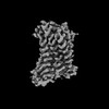 36901MC 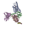 8i7vC 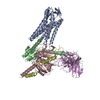 8i7wC 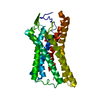 8k5bC 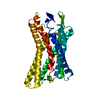 8k5dC M: map data used to model this data C: citing same article ( |
|---|---|
| Similar structure data |
-Links
 PDBj
PDBj


- Assembly
| Deposited unit | 
|
|---|---|
| 1 |
|
-Components
| #1: Protein | Mass: 54320.297 Da / Num. of mol.: 1 Source method: isolated from a genetically manipulated source Source: (gene. exp.) Homo sapiens (human) / Gene: HCAR2, HCA2 / Production host:   Spodoptera frugiperda (fall armyworm) / References: UniProt: Q8TDS4 Spodoptera frugiperda (fall armyworm) / References: UniProt: Q8TDS4 |
|---|---|
| #2: Chemical | ChemComp-OJX /   Mass: 155.131 Da / Num. of mol.: 1 / Source method: obtained synthetically / Formula: C6H7N2O3 / Feature type: SUBJECT OF INVESTIGATION Mass: 155.131 Da / Num. of mol.: 1 / Source method: obtained synthetically / Formula: C6H7N2O3 / Feature type: SUBJECT OF INVESTIGATION |
| Has ligand of interest | Y |
-Experimental details
-Experiment
| Experiment | Method: ELECTRON MICROSCOPY |
|---|---|
| EM experiment | Aggregation state: PARTICLE / 3D reconstruction method: single particle reconstruction |
- Sample preparation
| Component | Name: Acipimox bound human hydroxy-carboxylic acid receptor 2 Type: COMPLEX / Entity ID: #1 / Source: RECOMBINANT |
|---|---|
| Molecular weight | Value: 0.15 MDa / Experimental value: YES |
| Source (natural) | Organism: Homo sapiens (human) |
| Source (recombinant) | Organism: Spodoptera frugiperda (fall armyworm) |
| Buffer solution | pH: 7.5 Details: 20 mM HEPES pH8.0, 100 mM NaCl, 1 mM MgCl2, 0.5 mM TCEP, 0.001% LMNG, 0.0001% CHS |
| Specimen | Embedding applied: NO / Shadowing applied: NO / Staining applied: NO / Vitrification applied: YES |
| Specimen support | Grid material: COPPER / Grid mesh size: 300 divisions/in. / Grid type: Quantifoil R1.2/1.3 |
| Vitrification | Instrument: FEI VITROBOT MARK IV / Cryogen name: ETHANE / Humidity: 100 % / Chamber temperature: 277 K |
- Electron microscopy imaging
| Experimental equipment |  Model: Titan Krios / Image courtesy: FEI Company |
|---|---|
| Microscopy | Model: FEI TITAN KRIOS |
| Electron gun | Electron source: FIELD EMISSION GUN / Accelerating voltage: 300 kV / Illumination mode: FLOOD BEAM |
| Electron lens | Mode: BRIGHT FIELDBright-field microscopy / Nominal magnification: 105000 X / Nominal defocus max: 1800 nm / Nominal defocus min: 800 nm / Cs: 2.7 mm |
| Specimen holder | Cryogen: NITROGEN / Specimen holder model: FEI TITAN KRIOS AUTOGRID HOLDER |
| Image recording | Electron dose: 50 e/Å2 / Film or detector model: GATAN K3 (6k x 4k) |
- Processing
| EM software |
| |||||||||||||||||||||||||||||||||
|---|---|---|---|---|---|---|---|---|---|---|---|---|---|---|---|---|---|---|---|---|---|---|---|---|---|---|---|---|---|---|---|---|---|---|
| CTF correction | Type: PHASE FLIPPING AND AMPLITUDE CORRECTION | |||||||||||||||||||||||||||||||||
| Symmetry | Point symmetry: C1 (asymmetric) | |||||||||||||||||||||||||||||||||
| 3D reconstruction | Resolution: 3.13 Å / Resolution method: FSC 0.143 CUT-OFF / Num. of particles: 228127 / Symmetry type: POINT | |||||||||||||||||||||||||||||||||
| Atomic model building | Protocol: AB INITIO MODEL / Space: REAL |
