 Movie
Movie Controller

[English] 日本語
 Yorodumi
- PDB-8iei: Cryo-EM structure of GPR156A/B of G-protein free GPR156 (local refine) -
+ Open data

- Basic information
| Entry | Database: PDB / ID: 8iei | |||||||||
|---|---|---|---|---|---|---|---|---|---|---|
| Title | Cryo-EM structure of GPR156A/B of G-protein free GPR156 (local refine) | |||||||||
 Components Components | Probable G-protein coupled receptor 156 | |||||||||
 Keywords Keywords |  MEMBRANE PROTEIN / G-protein coupled receptor / Signal transduction / Phospholipid MEMBRANE PROTEIN / G-protein coupled receptor / Signal transduction / Phospholipid | |||||||||
| Function / homology |  Function and homology information Function and homology informationG protein-coupled GABA receptor activity / G protein-coupled receptor heterodimeric complex / gamma-aminobutyric acid signaling pathway / plasma membraneSimilarity search - Function | |||||||||
| Biological species |  Homo sapiens (human) Homo sapiens (human) | |||||||||
| Method | ELECTRON MICROSCOPY / single particle reconstruction / cryo EM / Resolution: 2.62 Å | |||||||||
 Authors Authors | Shin, J. / Park, J. / Cho, Y. | |||||||||
| Funding support |  Korea, Republic Of, 1items Korea, Republic Of, 1items
| |||||||||
 Citation Citation | Journal: Nat Struct Mol Biol / Year: 2024 Title: Constitutive activation mechanism of a class C GPCR. Authors: Jinwoo Shin / Junhyeon Park / Jieun Jeong / Jordy Homing Lam / Xingyu Qiu / Di Wu / Kuglae Kim / Joo-Youn Lee / Carol V Robinson / Jaekyung Hyun / Vsevolod Katritch / Kwang Pyo Kim / Yunje Cho /   Abstract: Class C G-protein-coupled receptors (GPCRs) are activated through binding of agonists to the large extracellular domain (ECD) followed by rearrangement of the transmembrane domains (TMDs). GPR156, a ...Class C G-protein-coupled receptors (GPCRs) are activated through binding of agonists to the large extracellular domain (ECD) followed by rearrangement of the transmembrane domains (TMDs). GPR156, a class C orphan GPCR, is unique because it lacks an ECD and exhibits constitutive activity. Impaired GPR156-G signaling contributes to loss of hearing. Here we present the cryo-electron microscopy structures of human GPR156 in the G-free and G-coupled states. We found that an endogenous phospholipid molecule is located within each TMD of the GPR156 dimer. Asymmetric binding of Gα to the phospholipid-bound GPR156 dimer restructures the first and second intracellular loops and the carboxy-terminal part of the elongated transmembrane 7 (TM7) without altering dimer conformation. Our findings reveal that GPR156 is a transducer for phospholipid signaling. Constant binding of abundant phospholipid molecules and the G-protein-induced reshaping of the cytoplasmic face provide a basis for the constitutive activation of GPR156. | |||||||||
| History |
|
- Structure visualization
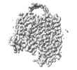
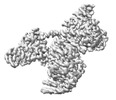
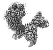
| Structure viewer | Molecule: MolmilJmol/JSmol |
|---|
- Downloads & links
-Download
| PDBx/mmCIF format | 8iei.cif.gz | 132.4 KB | Display | PDBx/mmCIF format |
|---|---|---|---|---|
| PDB format | pdb8iei.ent.gz | Display | PDB format | |
| PDBx/mmJSON format | 8iei.json.gz | Tree view | PDBx/mmJSON format | |
| Others |  Other downloads Other downloads |
-Validation report
| Arichive directory | https://data.pdbj.org/pub/pdb/validation_reports/ie/8ieiftp://data.pdbj.org/pub/pdb/validation_reports/ie/8iei | HTTPS FTP |
|---|
-Related structure data
| Related structure data |  35382MC 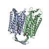 8iebC 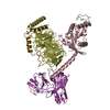 8iecC  8iedC 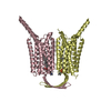 8iepC 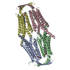 8ieqC M: map data used to model this data C: citing same article ( |
|---|---|
| Similar structure data |
-Links
 PDBj
PDBj
- Assembly
| Deposited unit | 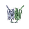
|
|---|---|
| 1 |
|
-Components
| #1: Protein | Mass: 65628.484 Da / Num. of mol.: 2 Source method: isolated from a genetically manipulated source Source: (gene. exp.) Homo sapiens (human) / Gene: GPR156 / Cell line (production host): HEK293 / Production host: Homo sapiens (human) / References: UniProt: Q8NFN8#2: Chemical | Mass: 760.076 Da / Num. of mol.: 2 / Source method: obtained synthetically / Formula: C42H82NO8P / Feature type: SUBJECT OF INVESTIGATION Has ligand of interest | Y | |
|---|
-Experimental details
-Experiment
| Experiment | Method: ELECTRON MICROSCOPY |
|---|---|
| EM experiment | Aggregation state: PARTICLE / 3D reconstruction method: single particle reconstruction |
- Sample preparation
| Component | Name: GPR156A/B / Type: COMPLEX / Entity ID: #1 / Source: RECOMBINANT |
|---|---|
| Molecular weight | Value: 131.2 kDa/nm / Experimental value: NO |
| Source (natural) | Organism: Homo sapiens (human) |
| Source (recombinant) | Organism: Homo sapiens (human) |
| Buffer solution | pH: 7.5 |
| Specimen | Embedding applied: NO / Shadowing applied: NO / Staining applied: NO / Vitrification applied: YES |
| Vitrification | Cryogen name: ETHANE |
- Electron microscopy imaging
| Experimental equipment |  Model: Titan Krios / Image courtesy: FEI Company |
|---|---|
| Microscopy | Model: FEI TITAN KRIOS |
| Electron gun | Electron source: OTHER / Accelerating voltage: 300 kV / Illumination mode: OTHER |
| Electron lens | Mode: OTHER / Nominal defocus max: 2000 nm / Nominal defocus min: 1000 nm |
| Image recording | Electron dose: 64 e/Å2 / Film or detector model: FEI FALCON IV (4k x 4k) |
- Processing
| CTF correction | Type: PHASE FLIPPING AND AMPLITUDE CORRECTION | ||||||||||||||||||||||||
|---|---|---|---|---|---|---|---|---|---|---|---|---|---|---|---|---|---|---|---|---|---|---|---|---|---|
| 3D reconstruction | Resolution: 2.62 Å / Resolution method: FSC 0.143 CUT-OFF / Num. of particles: 493410 / Symmetry type: POINT | ||||||||||||||||||||||||
| Refine LS restraints |
|
