 Movie
Movie Controller


+ Open data

- Basic information
| Entry | Database: PDB / ID: 8g42 | |||||||||
|---|---|---|---|---|---|---|---|---|---|---|
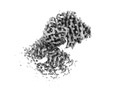
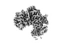
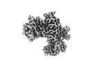
| Title | KRAS G12C complex with GDP imaged on a cryo-EM imaging scaffold | |||||||||
 Components Components |
| |||||||||
 Keywords Keywords |  SIGNALING PROTEIN / CryoEM imaging scaffold / Cancer / GTPase SIGNALING PROTEIN / CryoEM imaging scaffold / Cancer / GTPase | |||||||||
| Function / homology |  Function and homology information Function and homology informationforebrain astrocyte development / negative regulation of epithelial cell differentiation / regulation of synaptic transmission, GABAergic / epithelial tube branching involved in lung morphogenesis / type I pneumocyte differentiation / Rac protein signal transduction / skeletal muscle cell differentiation / positive regulation of Rac protein signal transduction / Signaling by RAS GAP mutants / Signaling by RAS GTPase mutants ...forebrain astrocyte development / negative regulation of epithelial cell differentiation / regulation of synaptic transmission, GABAergic / epithelial tube branching involved in lung morphogenesis / type I pneumocyte differentiation / Rac protein signal transduction / skeletal muscle cell differentiation / positive regulation of Rac protein signal transduction / Signaling by RAS GAP mutants / Signaling by RAS GTPase mutants / Activation of RAS in B cells / RAS signaling downstream of NF1 loss-of-function variants / RUNX3 regulates p14-ARF / SOS-mediated signalling / Activated NTRK3 signals through RAS / Activated NTRK2 signals through RAS / SHC1 events in ERBB4 signaling / Signalling to RAS / glial cell proliferation / SHC-related events triggered by IGF1R / Activated NTRK2 signals through FRS2 and FRS3 / Estrogen-stimulated signaling through PRKCZ / SHC-mediated cascade:FGFR3 / MET activates RAS signaling / PTK6 Regulates RHO GTPases, RAS GTPase and MAP kinases / Signaling by PDGFRA transmembrane, juxtamembrane and kinase domain mutants / Signaling by PDGFRA extracellular domain mutants / SHC-mediated cascade:FGFR2 / SHC-mediated cascade:FGFR4 / Signaling by FGFR4 in disease / SHC-mediated cascade:FGFR1 / Erythropoietin activates RAS / protein-membrane adaptor activity / FRS-mediated FGFR3 signaling / Signaling by CSF3 (G-CSF) / positive regulation of glial cell proliferation / Signaling by FLT3 ITD and TKD mutants / homeostasis of number of cells within a tissue / FRS-mediated FGFR2 signaling / FRS-mediated FGFR4 signaling / Signaling by FGFR3 in disease / FRS-mediated FGFR1 signaling / p38MAPK events / Tie2 Signaling / Signaling by FGFR2 in disease / striated muscle cell differentiation / GRB2 events in EGFR signaling / SHC1 events in EGFR signaling / EGFR Transactivation by Gastrin / Signaling by FLT3 fusion proteins / FLT3 Signaling / Signaling by FGFR1 in disease / Ras activation upon Ca2+ influx through NMDA receptor / GRB2 events in ERBB2 signaling / NCAM signaling for neurite out-growth / CD209 (DC-SIGN) signaling / SHC1 events in ERBB2 signaling / Downstream signal transduction / Constitutive Signaling by Overexpressed ERBB2 / Insulin receptor signalling cascade / small monomeric GTPase / G protein activity / Signaling by phosphorylated juxtamembrane, extracellular and kinase domain KIT mutants / VEGFR2 mediated cell proliferation / FCERI mediated MAPK activation / Signaling by ERBB2 TMD/JMD mutants / regulation of long-term neuronal synaptic plasticity / RAF activation / Signaling by high-kinase activity BRAF mutants / Constitutive Signaling by EGFRvIII / visual learning / MAP2K and MAPK activation / Signaling by ERBB2 ECD mutants / Signaling by ERBB2 KD Mutants / Signaling by SCF-KIT / cytoplasmic side of plasma membrane / Regulation of RAS by GAPs / Negative regulation of MAPK pathway / RAS processing / Signaling by RAF1 mutants / GDP binding / Signaling by moderate kinase activity BRAF mutants / Paradoxical activation of RAF signaling by kinase inactive BRAF / Signaling downstream of RAS mutants / Signaling by CSF1 (M-CSF) in myeloid cells / MAPK cascade / Signaling by BRAF and RAF1 fusions / DAP12 signaling / Constitutive Signaling by Ligand-Responsive EGFR Cancer Variants / Ca2+ pathway / gene expression / actin cytoskeleton organization / RAF/MAP kinase cascade / neuron apoptotic process / mitochondrial outer membrane / negative regulation of neuron apoptotic process / Ras protein signal transduction / positive regulation of protein phosphorylation / Golgi membrane / focal adhesionSimilarity search - Function | |||||||||
| Biological species | synthetic construct (others) Homo sapiens (human) Homo sapiens (human) | |||||||||
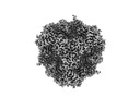
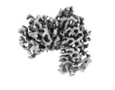
| Method | ELECTRON MICROSCOPY / single particle reconstruction / cryo EM / Resolution: 3.02 Å | |||||||||
 Authors Authors | Castells-Graells, R. / Sawaya, M.R. / Yeates, T.O. | |||||||||
| Funding support |  United States, 2items United States, 2items
| |||||||||
 Citation Citation | Journal: Proc Natl Acad Sci U S A / Year: 2023 Title: Cryo-EM structure determination of small therapeutic protein targets at 3 Å-resolution using a rigid imaging scaffold. Authors: Roger Castells-Graells / Kyle Meador / Mark A Arbing / Michael R Sawaya / Morgan Gee / Duilio Cascio / Emma Gleave / Judit É Debreczeni / Jason Breed / Karoline Leopold / Ankoor Patel / ...Authors: Roger Castells-Graells / Kyle Meador / Mark A Arbing / Michael R Sawaya / Morgan Gee / Duilio Cascio / Emma Gleave / Judit É Debreczeni / Jason Breed / Karoline Leopold / Ankoor Patel / Dushyant Jahagirdar / Bronwyn Lyons / Sriram Subramaniam / Chris Phillips / Todd O Yeates /   Abstract: Cryoelectron microscopy (Cryo-EM) has enabled structural determination of proteins larger than about 50 kDa, including many intractable by any other method, but it has largely failed for smaller ...Cryoelectron microscopy (Cryo-EM) has enabled structural determination of proteins larger than about 50 kDa, including many intractable by any other method, but it has largely failed for smaller proteins. Here, we obtain structures of small proteins by binding them to a rigid molecular scaffold based on a designed protein cage, revealing atomic details at resolutions reaching 2.9 Å. We apply this system to the key cancer signaling protein KRAS (19 kDa in size), obtaining four structures of oncogenic mutational variants by cryo-EM. Importantly, a structure for the key G12C mutant bound to an inhibitor drug (AMG510) reveals significant conformational differences compared to prior data in the crystalline state. The findings highlight the promise of cryo-EM scaffolds for advancing the design of drug molecules against small therapeutic protein targets in cancer and other human diseases. | |||||||||
| History |
|
- Structure visualization
| Structure viewer | Molecule: MolmilJmol/JSmol |
|---|
- Downloads & links
-Download
| PDBx/mmCIF format | 8g42.cif.gz | 97.5 KB | Display | PDBx/mmCIF format |
|---|---|---|---|---|
| PDB format | pdb8g42.ent.gz | 55.7 KB | Display | PDB format |
| PDBx/mmJSON format | 8g42.json.gz | Tree view | PDBx/mmJSON format | |
| Others |  Other downloads Other downloads |
-Validation report
| Arichive directory | https://data.pdbj.org/pub/pdb/validation_reports/g4/8g42ftp://data.pdbj.org/pub/pdb/validation_reports/g4/8g42 | HTTPS FTP |
|---|
-Related structure data
| Related structure data | 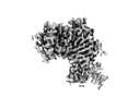 29713MC 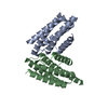 8g3kC 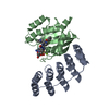 8g47C 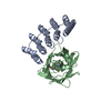 8g4eC 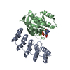 8g4fC 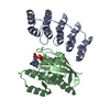 8g4hC M: map data used to model this data C: citing same article ( |
|---|---|
| Similar structure data |
-Links
 PDBj
PDBj


- Assembly
| Deposited unit | 
|
|---|---|
| 1 |
|
-Components
| #1: Protein | Mass: 35452.441 Da / Num. of mol.: 1 Source method: isolated from a genetically manipulated source Source: (gene. exp.) synthetic construct (others) / Production host:  Escherichia coli (E. coli) Escherichia coli (E. coli) |
|---|---|
| #2: Protein | Mass: 21622.275 Da / Num. of mol.: 1 / Mutation: G12C Source method: isolated from a genetically manipulated source Source: (gene. exp.) Homo sapiens (human) / Gene: KRAS, KRAS2, RASK2 / Production host: Escherichia coli (E. coli) / References: UniProt: P01116, small monomeric GTPase |
| #3: Chemical | ChemComp-GDP / Guanosine diphosphate  Type: RNA linking / Mass: 443.201 Da / Num. of mol.: 1 / Source method: obtained synthetically / Formula: C10H15N5O11P2 / Comment: GDP, energy-carrying molecule*YM Type: RNA linking / Mass: 443.201 Da / Num. of mol.: 1 / Source method: obtained synthetically / Formula: C10H15N5O11P2 / Comment: GDP, energy-carrying molecule*YM |
| #4: Chemical | ChemComp-MG /   Mass: 24.305 Da / Num. of mol.: 1 / Source method: obtained synthetically / Formula: Mg Mass: 24.305 Da / Num. of mol.: 1 / Source method: obtained synthetically / Formula: Mg |
| Has ligand of interest | N |
-Experimental details
-Experiment
| Experiment | Method: ELECTRON MICROSCOPY |
|---|---|
| EM experiment | Aggregation state: PARTICLE / 3D reconstruction method: single particle reconstruction |
- Sample preparation
| Component | Name: KRAS G12C displayed on a Cryo-EM imaging scaffold / Type: COMPLEX / Entity ID: #1-#2 / Source: RECOMBINANT |
|---|---|
| Molecular weight | Experimental value: NO |
| Source (natural) | Organism: synthetic construct (others) |
| Source (recombinant) | Organism: Escherichia coli (E. coli) |
| Buffer solution | pH: 8 |
| Specimen | Embedding applied: NO / Shadowing applied: NO / Staining applied: NO / Vitrification applied: YES |
| Specimen support | Grid material: COPPER / Grid mesh size: 300 divisions/in. / Grid type: Quantifoil R2/2 |
| Vitrification | Instrument: FEI VITROBOT MARK IV / Cryogen name: ETHANE |
- Electron microscopy imaging
| Experimental equipment |  Model: Titan Krios / Image courtesy: FEI Company |
|---|---|
| Microscopy | Model: FEI TITAN KRIOS |
| Electron gun | Electron source: FIELD EMISSION GUN / Accelerating voltage: 300 kV / Illumination mode: FLOOD BEAM |
| Electron lens | Mode: BRIGHT FIELDBright-field microscopy / Nominal defocus max: 2250 nm / Nominal defocus min: 1000 nm / Cs: 2.7 mm |
| Image recording | Electron dose: 40 e/Å2 / Film or detector model: FEI FALCON IV (4k x 4k) |
- Processing
| Software |
| ||||||||||||||||||||||||
|---|---|---|---|---|---|---|---|---|---|---|---|---|---|---|---|---|---|---|---|---|---|---|---|---|---|
| EM software |
| ||||||||||||||||||||||||
| CTF correction | Type: PHASE FLIPPING AND AMPLITUDE CORRECTION | ||||||||||||||||||||||||
| Symmetry | Point symmetry: C1 (asymmetric) | ||||||||||||||||||||||||
| 3D reconstruction | Resolution: 3.02 Å / Resolution method: FSC 0.143 CUT-OFF / Num. of particles: 189266 / Symmetry type: POINT | ||||||||||||||||||||||||
| Atomic model building | Protocol: AB INITIO MODEL / Space: REAL | ||||||||||||||||||||||||
| Refinement | Cross valid method: NONE Stereochemistry target values: GeoStd + Monomer Library + CDL v1.2 | ||||||||||||||||||||||||
| Displacement parameters | Biso mean: 48.32 Å2 | ||||||||||||||||||||||||
| Refine LS restraints |
|
