 Movie
Movie Controller


+ Open data

- Basic information
| Entry | Database: PDB / ID: 8f86 | ||||||
|---|---|---|---|---|---|---|---|
| Title | SIRT6 bound to an H3K9Ac nucleosome | ||||||
 Components Components |
| ||||||
 Keywords Keywords |  GENE REGULATION / nucleosome / SIRTUIN6 / histone deacylation / H3K9Ac GENE REGULATION / nucleosome / SIRTUIN6 / histone deacylation / H3K9Ac | ||||||
| Function / homology |  Function and homology information Function and homology informationNAD-dependent histone H3K56 deacetylase activity / NAD-dependent histone H3K18 deacetylase activity / ketone biosynthetic process / NAD-dependent histone H3K9 deacetylase activity / protein delipidation / regulation of lipid catabolic process / NAD+- protein-lysine ADP-ribosyltransferase activity / NAD-dependent protein demyristoylase activity / NAD-dependent protein depalmitoylase activity / chromosome, subtelomeric region ...NAD-dependent histone H3K56 deacetylase activity / NAD-dependent histone H3K18 deacetylase activity / ketone biosynthetic process / NAD-dependent histone H3K9 deacetylase activity / protein delipidation / regulation of lipid catabolic process / NAD+- protein-lysine ADP-ribosyltransferase activity / NAD-dependent protein demyristoylase activity / NAD-dependent protein depalmitoylase activity / chromosome, subtelomeric region / pericentric heterochromatin formation / NAD+-protein-arginine ADP-ribosyltransferase activity / positive regulation of protein localization to chromatin / DNA damage sensor activity / positive regulation of stem cell differentiation / positive regulation of blood vessel branching / retrotransposon silencing / NAD-dependent protein lysine deacetylase activity / protein localization to site of double-strand break / cardiac muscle cell differentiation / protein acetyllysine N-acetyltransferase / NAD-dependent histone deacetylase activity / positive regulation of chondrocyte proliferation / positive regulation of telomere maintenance / protein deacetylation / negative regulation of glucose import / TORC2 complex binding / lncRNA binding / negative regulation of glycolytic process / DNA repair-dependent chromatin remodeling / negative regulation of protein localization to chromatin / positive regulation of double-strand break repair / positive regulation of vascular endothelial cell proliferation / negative regulation of gene expression, epigenetic / regulation of double-strand break repair via homologous recombination / negative regulation of protein import into nucleus / positive regulation of stem cell population maintenance / positive regulation of stem cell proliferation / regulation of protein secretion / negative regulation of transcription elongation by RNA polymerase II / site of DNA damage / NAD+-protein ADP-ribosyltransferase activity / negative regulation of cellular senescence / subtelomeric heterochromatin formation / regulation of lipid metabolic process / NAD+ ADP-ribosyltransferase activity / Transferases; Glycosyltransferases; Pentosyltransferases / nucleosome binding / NAD+ binding / positive regulation of fat cell differentiation / negative regulation of gluconeogenesis / regulation of protein localization to plasma membrane / pericentric heterochromatin / response to UV / Transferases; Acyltransferases; Transferring groups other than aminoacyl groups / nucleotidyltransferase activity / positive regulation of protein export from nucleus / determination of adult lifespan / base-excision repair / circadian regulation of gene expression / protein destabilization / regulation of circadian rhythm / positive regulation of insulin secretion / chromatin DNA binding / Pre-NOTCH Transcription and Translation / structural constituent of chromatin / transcription corepressor activity / double-strand break repair / positive regulation of fibroblast proliferation / nucleosome / positive regulation of proteasomal ubiquitin-dependent protein catabolic process / glucose homeostasis / site of double-strand break / positive regulation of cold-induced thermogenesis / Processing of DNA double-strand break ends / damaged DNA binding / chromatin remodeling / protein heterodimerization activity / negative regulation of cell population proliferation / intracellular membrane-bounded organelle / chromatin binding / chromatin / negative regulation of transcription by RNA polymerase II / endoplasmic reticulum / protein homodimerization activity / DNA binding / zinc ion binding / nucleoplasm / nucleusSimilarity search - Function | ||||||
| Biological species | Xenopus laevis (African clawed frog) synthetic construct (others)  Homo sapiens (human) Homo sapiens (human) | ||||||
| Method | ELECTRON MICROSCOPY / single particle reconstruction / cryo EM / Resolution: 3.1 Å | ||||||
 Authors Authors | Markert, J. / Whedon, S. / Wang, Z. / Cole, P. / Farnung, L. | ||||||
| Funding support | 1items
| ||||||
 Citation Citation | Journal: J Am Chem Soc / Year: 2023 Title: Structural Basis of Sirtuin 6-Catalyzed Nucleosome Deacetylation. Authors: Zhipeng A Wang / Jonathan W Markert / Samuel D Whedon / Maheeshi Yapa Abeywardana / Kwangwoon Lee / Hanjie Jiang / Carolay Suarez / Hening Lin / Lucas Farnung / Philip A Cole /  Abstract: The reversible acetylation of histone lysine residues is controlled by the action of acetyltransferases and deacetylases (HDACs), which regulate chromatin structure and gene expression. The sirtuins ...The reversible acetylation of histone lysine residues is controlled by the action of acetyltransferases and deacetylases (HDACs), which regulate chromatin structure and gene expression. The sirtuins are a family of NAD-dependent HDAC enzymes, and one member, sirtuin 6 (Sirt6), influences DNA repair, transcription, and aging. Here, we demonstrate that Sirt6 is efficient at deacetylating several histone H3 acetylation sites, including its canonical site Lys9, in the context of nucleosomes but not free acetylated histone H3 protein substrates. By installing a chemical warhead at the Lys9 position of histone H3, we trap a catalytically poised Sirt6 in complex with a nucleosome and employ this in cryo-EM structural analysis. The structure of Sirt6 bound to a nucleosome reveals extensive interactions between distinct segments of Sirt6 and the H2A/H2B acidic patch and nucleosomal DNA, which accounts for the rapid deacetylation of nucleosomal H3 sites and the disfavoring of histone H2B acetylation sites. These findings provide a new framework for understanding how HDACs target and regulate chromatin. | ||||||
| History |
|
- Structure visualization
| Structure viewer | Molecule: MolmilJmol/JSmol |
|---|
- Downloads & links
-Download
| PDBx/mmCIF format | 8f86.cif.gz | 382.2 KB | Display | PDBx/mmCIF format |
|---|---|---|---|---|
| PDB format | pdb8f86.ent.gz | 290.2 KB | Display | PDB format |
| PDBx/mmJSON format | 8f86.json.gz | Tree view | PDBx/mmJSON format | |
| Others |  Other downloads Other downloads |
-Validation report
| Arichive directory | https://data.pdbj.org/pub/pdb/validation_reports/f8/8f86ftp://data.pdbj.org/pub/pdb/validation_reports/f8/8f86 | HTTPS FTP |
|---|
-Related structure data
| Related structure data | 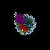 28915MC M: map data used to model this data C: citing same article ( |
|---|---|
| Similar structure data |
-Links
 PDBj
PDBj


- Assembly
| Deposited unit | 
|
|---|---|
| 1 |
|
-Components
-Protein , 5 types, 9 molecules AEBFCGDHK
| #1: Protein | Mass: 15271.863 Da / Num. of mol.: 2 Source method: isolated from a genetically manipulated source Source: (gene. exp.) Xenopus laevis (African clawed frog) / Production host:  Escherichia coli (E. coli) / References: UniProt: P84233 Escherichia coli (E. coli) / References: UniProt: P84233#2: Protein | Mass: 11263.231 Da / Num. of mol.: 2 Source method: isolated from a genetically manipulated source Source: (gene. exp.) Xenopus laevis (African clawed frog) / Production host: Escherichia coli (E. coli) / References: UniProt: P62799#3: Protein | Mass: 13978.241 Da / Num. of mol.: 2 Source method: isolated from a genetically manipulated source Source: (gene. exp.) Xenopus laevis (African clawed frog) / Production host: Escherichia coli (E. coli) / References: UniProt: P06897#4: Protein | / H2B1.1Mass: 13524.752 Da / Num. of mol.: 2 Source method: isolated from a genetically manipulated source Source: (gene. exp.) Xenopus laevis (African clawed frog) / Production host: Escherichia coli (E. coli) / References: UniProt: P02281#7: Protein | | Mass: 39152.863 Da / Num. of mol.: 1 Source method: isolated from a genetically manipulated source Source: (gene. exp.) Homo sapiens (human) / Gene: SIRT6, SIR2L6 / Production host: Escherichia coli (E. coli)References: UniProt: Q8N6T7, Transferases; Acyltransferases; Transferring groups other than aminoacyl groups, protein acetyllysine N-acetyltransferase, Transferases; Glycosyltransferases; Pentosyltransferases |
|---|
-DNA chain , 2 types, 2 molecules IJ
| #5: DNA chain | Mass: 57400.559 Da / Num. of mol.: 1 Source method: isolated from a genetically manipulated source Source: (gene. exp.) synthetic construct (others) / Production host: Escherichia coli (E. coli) |
|---|---|
| #6: DNA chain | Mass: 56831.191 Da / Num. of mol.: 1 Source method: isolated from a genetically manipulated source Source: (gene. exp.) synthetic construct (others) / Production host: Escherichia coli (E. coli) |
-Non-polymers , 2 types, 2 molecules 

| #8: Chemical | ChemComp-ZSL / [( Mass: 587.392 Da / Num. of mol.: 1 / Source method: obtained synthetically / Formula: C16H23N5O13P2S / Feature type: SUBJECT OF INVESTIGATION Mass: 587.392 Da / Num. of mol.: 1 / Source method: obtained synthetically / Formula: C16H23N5O13P2S / Feature type: SUBJECT OF INVESTIGATION |
|---|---|
| #9: Chemical | ChemComp-ZN /  Mass: 65.409 Da / Num. of mol.: 1 / Source method: obtained synthetically / Formula: Zn / Feature type: SUBJECT OF INVESTIGATION Mass: 65.409 Da / Num. of mol.: 1 / Source method: obtained synthetically / Formula: Zn / Feature type: SUBJECT OF INVESTIGATION |
-Details
| Has ligand of interest | Y |
|---|
-Experimental details
-Experiment
| Experiment | Method: ELECTRON MICROSCOPY |
|---|---|
| EM experiment | Aggregation state: PARTICLE / 3D reconstruction method: single particle reconstruction |
- Sample preparation
| Component | Name: SIRT6 bound to H3K9Ac / Type: COMPLEX / Entity ID: #1-#7 / Source: MULTIPLE SOURCES |
|---|---|
| Molecular weight | Experimental value: NO |
| Buffer solution | pH: 7.5 |
| Specimen | Embedding applied: NO / Shadowing applied: NO / Staining applied: NO / Vitrification applied: YES |
| Vitrification | Cryogen name: ETHANE |
- Electron microscopy imaging
| Experimental equipment |  Model: Titan Krios / Image courtesy: FEI Company |
|---|---|
| Microscopy | Model: FEI TITAN KRIOS |
| Electron gun | Electron source: FIELD EMISSION GUN / Accelerating voltage: 300 kV / Illumination mode: OTHER |
| Electron lens | Mode: BRIGHT FIELDBright-field microscopy / Nominal defocus max: 1700 nm / Nominal defocus min: 700 nm |
| Image recording | Electron dose: 50.45 e/Å2 / Film or detector model: GATAN K3 BIOQUANTUM (6k x 4k) |
- Processing
| Software | Name: PHENIX / Version: 1.20.1_4487: / Classification: refinement | ||||||||||||||||||||||||
|---|---|---|---|---|---|---|---|---|---|---|---|---|---|---|---|---|---|---|---|---|---|---|---|---|---|
| CTF correction | Type: PHASE FLIPPING AND AMPLITUDE CORRECTION | ||||||||||||||||||||||||
| 3D reconstruction | Resolution: 3.1 Å / Resolution method: FSC 0.143 CUT-OFF / Num. of particles: 95205 / Symmetry type: POINT | ||||||||||||||||||||||||
| Atomic model building | Protocol: RIGID BODY FIT / Space: REAL | ||||||||||||||||||||||||
| Refine LS restraints |
|
