 Movie
Movie Controller

[English] 日本語
 Yorodumi
- PDB-8eyc: Crystal structure of PTP1B D181A/Q262A/C215A phosphatase domain w... -
+ Open data

- Basic information
| Entry | Database: PDB / ID: 8eyc | ||||||
|---|---|---|---|---|---|---|---|
| Title | Crystal structure of PTP1B D181A/Q262A/C215A phosphatase domain with TYK2 activation loop phosphopeptide | ||||||
 Components Components |
| ||||||
 Keywords Keywords |  SIGNALING PROTEIN / PTP1B / JAK/STAT / IRK SIGNALING PROTEIN / PTP1B / JAK/STAT / IRK | ||||||
| Function / homology |  Function and homology information Function and homology informationtype III interferon-mediated signaling pathway / interleukin-12 receptor complex / interleukin-23 receptor complex / Interleukin-23 signaling / positive regulation of T-helper 17 type immune response / type 1 angiotensin receptor binding / positive regulation of NK T cell proliferation / interleukin-12-mediated signaling pathway / Interleukin-12 signaling / Interleukin-35 Signalling ...type III interferon-mediated signaling pathway / interleukin-12 receptor complex / interleukin-23 receptor complex / Interleukin-23 signaling / positive regulation of T-helper 17 type immune response / type 1 angiotensin receptor binding / positive regulation of NK T cell proliferation / interleukin-12-mediated signaling pathway / Interleukin-12 signaling / Interleukin-35 Signalling / Interleukin-27 signaling / IL-6-type cytokine receptor ligand interactions / growth hormone receptor binding / positive regulation of natural killer cell proliferation / Other interleukin signaling / extrinsic component of plasma membrane / regulation of hepatocyte growth factor receptor signaling pathway / PTK6 Down-Regulation / Interleukin-20 family signaling / peptidyl-tyrosine dephosphorylation involved in inactivation of protein kinase activity / positive regulation of receptor catabolic process / insulin receptor recycling / type I interferon-mediated signaling pathway / Interleukin-6 signaling / negative regulation of PERK-mediated unfolded protein response / negative regulation of vascular endothelial growth factor receptor signaling pathway / regulation of intracellular protein transport / IRE1-mediated unfolded protein response / positive regulation of interleukin-17 production / MAPK3 (ERK1) activation / cytoplasmic side of endoplasmic reticulum membrane / platelet-derived growth factor receptor-beta signaling pathway / sorting endosome / mitochondrial crista / Interleukin-10 signaling / MAPK1 (ERK2) activation / positive regulation of IRE1-mediated unfolded protein response / cell surface receptor signaling pathway via JAK-STAT / regulation of type I interferon-mediated signaling pathway / regulation of endocytosis / non-membrane spanning protein tyrosine phosphatase activity / positive regulation of protein tyrosine kinase activity / peptidyl-tyrosine dephosphorylation / Regulation of IFNA/IFNB signaling / regulation of signal transduction / cellular response to unfolded protein / growth hormone receptor signaling pathway via JAK-STAT / negative regulation of endoplasmic reticulum stress-induced intrinsic apoptotic signaling pathway / negative regulation of signal transduction / type II interferon-mediated signaling pathway / Regulation of IFNG signaling / Growth hormone receptor signaling / MECP2 regulates neuronal receptors and channels / positive regulation of T cell proliferation / endoplasmic reticulum unfolded protein response / Signaling by CSF3 (G-CSF) / positive regulation of JUN kinase activity / Insulin receptor recycling / negative regulation of insulin receptor signaling pathway / ephrin receptor binding / Integrin signaling / protein dephosphorylation / protein-tyrosine-phosphatase / protein phosphatase 2A binding / negative regulation of MAP kinase activity / protein tyrosine phosphatase activity / Evasion by RSV of host interferon responses / endosome lumen / non-specific protein-tyrosine kinase / positive regulation of receptor signaling pathway via JAK-STAT / non-membrane spanning protein tyrosine kinase activity / Signaling by ALK fusions and activated point mutants / insulin receptor binding / Negative regulation of MET activity / Inactivation of CSF3 (G-CSF) signaling / negative regulation of ERK1 and ERK2 cascade / cytoplasmic side of plasma membrane / receptor tyrosine kinase binding / cellular response to virus / cytokine-mediated signaling pathway / positive regulation of protein localization to nucleus / positive regulation of type II interferon production / Interferon alpha/beta signaling / insulin receptor signaling pathway / actin cytoskeleton organization / protein tyrosine kinase activity / Interleukin-4 and Interleukin-13 signaling / Potential therapeutics for SARS / early endosome / cell differentiation / cytoskeleton / intracellular signal transduction / mitochondrial matrix / cadherin binding / immune response / protein phosphorylation / protein kinase binding / SARS-CoV-2 activates/modulates innate and adaptive immune responses / enzyme binding / endoplasmic reticulumSimilarity search - Function | ||||||
| Biological species |  Homo sapiens (human) Homo sapiens (human) | ||||||
| Method | X-RAY DIFFRACTION / SYNCHROTRON / MOLECULAR REPLACEMENT / Resolution: 2.99 Å | ||||||
 Authors Authors | Morris, R. / Kershaw, N.J. / Babon, J.J. | ||||||
| Funding support |  Australia, 1items Australia, 1items
| ||||||
 Citation Citation | Journal: Commun Biol / Year: 2023 Title: Structure guided studies of the interaction between PTP1B and JAK. Authors: Morris, R. / Keating, N. / Tan, C. / Chen, H. / Laktyushin, A. / Saiyed, T. / Liau, N.P.D. / Nicola, N.A. / Tiganis, T. / Kershaw, N.J. / Babon, J.J. | ||||||
| History |
|
- Structure visualization
| Structure viewer | Molecule: MolmilJmol/JSmol |
|---|
- Downloads & links
-Download
| PDBx/mmCIF format | 8eyc.cif.gz | 75.8 KB | Display | PDBx/mmCIF format |
|---|---|---|---|---|
| PDB format | pdb8eyc.ent.gz | 52.7 KB | Display | PDB format |
| PDBx/mmJSON format | 8eyc.json.gz | Tree view | PDBx/mmJSON format | |
| Others |  Other downloads Other downloads |
-Validation report
| Arichive directory | https://data.pdbj.org/pub/pdb/validation_reports/ey/8eycftp://data.pdbj.org/pub/pdb/validation_reports/ey/8eyc | HTTPS FTP |
|---|
-Related structure data
| Related structure data | 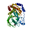 8exiC 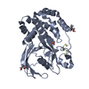 8exjC  8exkC  8exmC 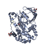 8exnC  8eyaC  8eybC  8f88C  4zrtS S: Starting model for refinement C: citing same article ( |
|---|---|
| Similar structure data |
-Links
 PDBj
PDBj


- Assembly
| Deposited unit | 
| ||||||||
|---|---|---|---|---|---|---|---|---|---|
| 1 |
| ||||||||
| Unit cell |
|
-Components
| #1: Protein | Mass: 34700.602 Da / Num. of mol.: 1 / Mutation: D181A/Q262A/C215A Source method: isolated from a genetically manipulated source Source: (gene. exp.) Homo sapiens (human) / Gene: PTPN1, PTP1B / Production host:  Escherichia coli (E. coli) / References: UniProt: P18031, protein-tyrosine-phosphatase Escherichia coli (E. coli) / References: UniProt: P18031, protein-tyrosine-phosphatase |
|---|---|
| #2: Protein/peptide | Mass: 1983.852 Da / Num. of mol.: 1 / Fragment: residues 1048-1062 of TYK2 / Source method: obtained synthetically / Source: (synth.) Homo sapiens (human)References: UniProt: P29597, non-specific protein-tyrosine kinase |
| #3: Water | ChemComp-HOH / Water Mass: 18.015 Da / Num. of mol.: 4 / Source method: isolated from a natural source / Formula: H2O Mass: 18.015 Da / Num. of mol.: 4 / Source method: isolated from a natural source / Formula: H2O |
| Has ligand of interest | Y |
-Experimental details
-Experiment
| Experiment | Method: X-RAY DIFFRACTION / Number of used crystals: 1 |
|---|
- Sample preparation
| Crystal | Density Matthews: 2.71 Å3/Da / Density % sol: 54.55 % |
|---|---|
| Crystal grow | Temperature: 281.15 K / Method: vapor diffusion, sitting drop Details: 14% PEG 8K, 0.20 M Magnesium Acetate, 0.1 M MES (pH 6.5) |
-Data collection
| Diffraction | Mean temperature: 100 K / Serial crystal experiment: N |
|---|---|
| Diffraction source | Source: SYNCHROTRON / Site: Australian Synchrotron / Beamline: MX2 / Wavelength: 0.95373 Å |
| Detector | Type: DECTRIS EIGER X 16M / Detector: PIXEL / Date: Jun 20, 2018 |
| Radiation | Protocol: SINGLE WAVELENGTH / Monochromatic (M) / Laue (L): M / Scattering type: x-ray |
| Radiation wavelength | Wavelength: 0.95373 Å / Relative weight: 1 |
| Reflection | Resolution: 2.79→48.861 Å / Num. obs: 64878 / % possible obs: 99.56 % / Redundancy: 6.5 % / CC1/2: 0.997 / Net I/σ(I): 10.44 |
| Reflection shell | Resolution: 2.79→2.89 Å / Num. unique obs: 6043 / CC1/2: 0.377 |
- Processing
| Software |
| |||||||||||||||||||||||||||||||||||||||||||||||||
|---|---|---|---|---|---|---|---|---|---|---|---|---|---|---|---|---|---|---|---|---|---|---|---|---|---|---|---|---|---|---|---|---|---|---|---|---|---|---|---|---|---|---|---|---|---|---|---|---|---|---|
| Refinement | Method to determine structure: MOLECULAR REPLACEMENT Starting model: 4ZRT Resolution: 2.99→48.86 Å / SU ML: 0.38 / Cross valid method: THROUGHOUT / σ(F): 1.35 / Phase error: 28.53 / Stereochemistry target values: ML
| |||||||||||||||||||||||||||||||||||||||||||||||||
| Solvent computation | Shrinkage radii: 0.9 Å / VDW probe radii: 1.1 Å / Solvent model: FLAT BULK SOLVENT MODEL | |||||||||||||||||||||||||||||||||||||||||||||||||
| Refinement step | Cycle: LAST / Resolution: 2.99→48.86 Å
| |||||||||||||||||||||||||||||||||||||||||||||||||
| Refine LS restraints |
| |||||||||||||||||||||||||||||||||||||||||||||||||
| LS refinement shell |
|
