 Movie
Movie Controller

[English] 日本語
 Yorodumi
- PDB-8ey0: Structure of an orthogonal PYR1*:HAB1* chemical-induced dimerizat... -
+ Open data

- Basic information
| Entry | Database: PDB / ID: 8ey0 | |||||||||
|---|---|---|---|---|---|---|---|---|---|---|
| Title | Structure of an orthogonal PYR1*:HAB1* chemical-induced dimerization module in complex with mandipropamid | |||||||||
 Components Components |
| |||||||||
 Keywords Keywords |  PLANT PROTEIN / PYR/PYL/RCAR / PYR1 / Hab1 / Hormone receptor / Biosensor PLANT PROTEIN / PYR/PYL/RCAR / PYR1 / Hab1 / Hormone receptor / Biosensor | |||||||||
| Function / homology |  Function and homology information Function and homology informationpositive regulation of response to water deprivation / plant-type vacuole membrane / regulation of protein serine/threonine phosphatase activity / protein phosphatase inhibitor complex / abscisic acid binding / abscisic acid-activated signaling pathway / protein phosphatase inhibitor activity / myosin phosphatase activity / protein serine/threonine phosphatase activity / protein-serine/threonine phosphatase ...positive regulation of response to water deprivation / plant-type vacuole membrane / regulation of protein serine/threonine phosphatase activity / protein phosphatase inhibitor complex / abscisic acid binding / abscisic acid-activated signaling pathway / protein phosphatase inhibitor activity / myosin phosphatase activity / protein serine/threonine phosphatase activity / protein-serine/threonine phosphatase / ubiquitin-like protein ligase binding / signaling receptor activity / protein homodimerization activity / identical protein binding / metal ion binding / nucleus / plasma membrane / cytosol / cytoplasmSimilarity search - Function | |||||||||
| Biological species |  Arabidopsis thaliana (thale cress) Arabidopsis thaliana (thale cress) | |||||||||
| Method | X-RAY DIFFRACTION / SYNCHROTRON / MOLECULAR REPLACEMENT / molecular replacement / Resolution: 2.4 Å | |||||||||
 Authors Authors | Park, S.-Y. / Volkman, B.F. / Cutler, S.R. / Peterson, F.C. | |||||||||
| Funding support |  United States, 2items United States, 2items
| |||||||||
 Citation Citation | Journal: Nat.Chem.Biol. / Year: 2024 Title: An orthogonalized PYR1-based CID module with reprogrammable ligand-binding specificity. Authors: Park, S.Y. / Qiu, J. / Wei, S. / Peterson, F.C. / Beltran, J. / Medina-Cucurella, A.V. / Vaidya, A.S. / Xing, Z. / Volkman, B.F. / Nusinow, D.A. / Whitehead, T.A. / Wheeldon, I. / Cutler, S.R. | |||||||||
| History |
|
- Structure visualization
| Structure viewer | Molecule: MolmilJmol/JSmol |
|---|
- Downloads & links
-Download
| PDBx/mmCIF format | 8ey0.cif.gz | 208 KB | Display | PDBx/mmCIF format |
|---|---|---|---|---|
| PDB format | pdb8ey0.ent.gz | 165.5 KB | Display | PDB format |
| PDBx/mmJSON format | 8ey0.json.gz | Tree view | PDBx/mmJSON format | |
| Others |  Other downloads Other downloads |
-Validation report
| Arichive directory | https://data.pdbj.org/pub/pdb/validation_reports/ey/8ey0ftp://data.pdbj.org/pub/pdb/validation_reports/ey/8ey0 | HTTPS FTP |
|---|
-Related structure data
| Related structure data | 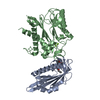 4wvoS S: Starting model for refinement |
|---|---|
| Similar structure data |
-Links
 PDBj
PDBj- Assembly
| Deposited unit | 
| ||||||||
|---|---|---|---|---|---|---|---|---|---|
| 1 |
| ||||||||
| Unit cell |
|
-Components
| #1: Protein | Mass: 36901.273 Da / Num. of mol.: 1 Source method: isolated from a genetically manipulated source Source: (gene. exp.) Arabidopsis thaliana (thale cress) / Gene: HAB1, P2C-HA, At1g72770, F28P22.4 / Plasmid: pET / Production host:  Escherichia coli BL21(DE3) (bacteria) Escherichia coli BL21(DE3) (bacteria)References: UniProt: Q9CAJ0, protein-serine/threonine phosphatase | ||||||
|---|---|---|---|---|---|---|---|
| #2: Protein | Mass: 20614.252 Da / Num. of mol.: 1 Source method: isolated from a genetically manipulated source Source: (gene. exp.) Arabidopsis thaliana (thale cress) / Gene: PYR1, ABIP6, RCAR11, At4g17870, T6K21.50 / Plasmid: pET / Production host: Escherichia coli BL21(DE3) (bacteria) / References: UniProt: O49686 | ||||||
| #3: Chemical | Glycerol  Mass: 92.094 Da / Num. of mol.: 2 / Source method: obtained synthetically / Formula: C3H8O3 Mass: 92.094 Da / Num. of mol.: 2 / Source method: obtained synthetically / Formula: C3H8O3#4: Chemical | ChemComp-3UZ / ( |   Mass: 411.878 Da / Num. of mol.: 1 / Source method: obtained synthetically / Formula: C23H22ClNO4 / Feature type: SUBJECT OF INVESTIGATION Mass: 411.878 Da / Num. of mol.: 1 / Source method: obtained synthetically / Formula: C23H22ClNO4 / Feature type: SUBJECT OF INVESTIGATION#5: Water | ChemComp-HOH / | Water Mass: 18.015 Da / Num. of mol.: 106 / Source method: isolated from a natural source / Formula: H2O Mass: 18.015 Da / Num. of mol.: 106 / Source method: isolated from a natural source / Formula: H2OHas ligand of interest | Y | |
-Experimental details
-Experiment
| Experiment | Method: X-RAY DIFFRACTION / Number of used crystals: 1 |
|---|
- Sample preparation
| Crystal | Density Matthews: 2.46 Å3/Da / Density % sol: 49.99 % |
|---|---|
| Crystal grow | Temperature: 292 K / Method: vapor diffusion, sitting drop / Details: 0.15 mM postassium bromide and 30% PEG 2K-MME |
-Data collection
| Diffraction | Mean temperature: 100 K / Serial crystal experiment: N |
|---|---|
| Diffraction source | Source: SYNCHROTRON / Site: APS / Beamline: 21-ID-G / Wavelength: 0.97857 Å |
| Detector | Type: MARMOSAIC 300 mm CCD / Detector: CCD / Date: Nov 14, 2015 |
| Radiation | Monochromator: C(111) / Protocol: SINGLE WAVELENGTH / Monochromatic (M) / Laue (L): M / Scattering type: x-ray |
| Radiation wavelength | Wavelength: 0.97857 Å / Relative weight: 1 |
| Reflection | Resolution: 2.4→50 Å / Num. obs: 21877 / % possible obs: 99.9 % / Redundancy: 7.5 % / Rmerge(I) obs: 0.073 / Rpim(I) all: 0.028 / Rrim(I) all: 0.078 / Net I/σ(I): 26.4 |
| Reflection shell | Resolution: 2.4→2.44 Å / Rmerge(I) obs: 0.847 / Mean I/σ(I) obs: 2.2 / Num. unique obs: 1126 / CC1/2: 0.782 / Rpim(I) all: 0.326 / Rrim(I) all: 0.909 / % possible all: 99.9 |
-Phasing
| Phasing | Method: molecular replacement | |||||||||
|---|---|---|---|---|---|---|---|---|---|---|
| Phasing MR |
|
- Processing
| Software |
| |||||||||||||||||||||||||||||||||||||||||||||||||||||||||||||||||||||||||||||||||||||||||||||||||||||||||||||||||||||||||||||||||||||||||||||||||||||||||||||||||||||||||||||||||||||||||||||||||||||||||||||||||||||||||||||||||||||||||||||||||||||||||||||||||||||||||||||||||||
|---|---|---|---|---|---|---|---|---|---|---|---|---|---|---|---|---|---|---|---|---|---|---|---|---|---|---|---|---|---|---|---|---|---|---|---|---|---|---|---|---|---|---|---|---|---|---|---|---|---|---|---|---|---|---|---|---|---|---|---|---|---|---|---|---|---|---|---|---|---|---|---|---|---|---|---|---|---|---|---|---|---|---|---|---|---|---|---|---|---|---|---|---|---|---|---|---|---|---|---|---|---|---|---|---|---|---|---|---|---|---|---|---|---|---|---|---|---|---|---|---|---|---|---|---|---|---|---|---|---|---|---|---|---|---|---|---|---|---|---|---|---|---|---|---|---|---|---|---|---|---|---|---|---|---|---|---|---|---|---|---|---|---|---|---|---|---|---|---|---|---|---|---|---|---|---|---|---|---|---|---|---|---|---|---|---|---|---|---|---|---|---|---|---|---|---|---|---|---|---|---|---|---|---|---|---|---|---|---|---|---|---|---|---|---|---|---|---|---|---|---|---|---|---|---|---|---|---|---|---|---|---|---|---|---|---|---|---|---|---|---|---|---|---|---|---|---|---|---|---|---|---|---|---|---|---|---|---|---|---|---|---|---|---|---|---|---|---|---|---|---|---|---|---|---|---|---|
| Refinement | Method to determine structure: MOLECULAR REPLACEMENT Starting model: 4wvo Resolution: 2.4→30.127 Å / SU ML: 0.31 / Cross valid method: THROUGHOUT / σ(F): 0 / Phase error: 24.36 / Stereochemistry target values: ML
| |||||||||||||||||||||||||||||||||||||||||||||||||||||||||||||||||||||||||||||||||||||||||||||||||||||||||||||||||||||||||||||||||||||||||||||||||||||||||||||||||||||||||||||||||||||||||||||||||||||||||||||||||||||||||||||||||||||||||||||||||||||||||||||||||||||||||||||||||||
| Solvent computation | Shrinkage radii: 0.9 Å / VDW probe radii: 1.11 Å / Solvent model: FLAT BULK SOLVENT MODEL | |||||||||||||||||||||||||||||||||||||||||||||||||||||||||||||||||||||||||||||||||||||||||||||||||||||||||||||||||||||||||||||||||||||||||||||||||||||||||||||||||||||||||||||||||||||||||||||||||||||||||||||||||||||||||||||||||||||||||||||||||||||||||||||||||||||||||||||||||||
| Displacement parameters | Biso max: 185.83 Å2 / Biso mean: 63.3957 Å2 / Biso min: 26.71 Å2 | |||||||||||||||||||||||||||||||||||||||||||||||||||||||||||||||||||||||||||||||||||||||||||||||||||||||||||||||||||||||||||||||||||||||||||||||||||||||||||||||||||||||||||||||||||||||||||||||||||||||||||||||||||||||||||||||||||||||||||||||||||||||||||||||||||||||||||||||||||
| Refinement step | Cycle: final / Resolution: 2.4→30.127 Å
| |||||||||||||||||||||||||||||||||||||||||||||||||||||||||||||||||||||||||||||||||||||||||||||||||||||||||||||||||||||||||||||||||||||||||||||||||||||||||||||||||||||||||||||||||||||||||||||||||||||||||||||||||||||||||||||||||||||||||||||||||||||||||||||||||||||||||||||||||||
| LS refinement shell | Refine-ID: X-RAY DIFFRACTION / Rfactor Rfree error: 0
| |||||||||||||||||||||||||||||||||||||||||||||||||||||||||||||||||||||||||||||||||||||||||||||||||||||||||||||||||||||||||||||||||||||||||||||||||||||||||||||||||||||||||||||||||||||||||||||||||||||||||||||||||||||||||||||||||||||||||||||||||||||||||||||||||||||||||||||||||||
| Refinement TLS params. | Method: refined / Refine-ID: X-RAY DIFFRACTION
| |||||||||||||||||||||||||||||||||||||||||||||||||||||||||||||||||||||||||||||||||||||||||||||||||||||||||||||||||||||||||||||||||||||||||||||||||||||||||||||||||||||||||||||||||||||||||||||||||||||||||||||||||||||||||||||||||||||||||||||||||||||||||||||||||||||||||||||||||||
| Refinement TLS group |
|
