 Movie
Movie Controller

[English] 日本語
 Yorodumi
- PDB-4wvo: An engineered PYR1 mandipropamid receptor in complex with mandipr... -
+ Open data

- Basic information
| Entry | Database: PDB / ID: 4wvo | ||||||
|---|---|---|---|---|---|---|---|
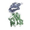
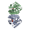
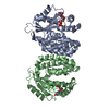
| Title | An engineered PYR1 mandipropamid receptor in complex with mandipropamid and HAB1 | ||||||
 Components Components |
| ||||||
 Keywords Keywords | HYDROLASE/HYDROLASE INTHIBITOR / PYR/PYL/RCAR /  PYR1 / Hab1 / mandipropamid / PP2C inhibitor / HYDROLASE-HYDROLASE INTHIBITOR complex PYR1 / Hab1 / mandipropamid / PP2C inhibitor / HYDROLASE-HYDROLASE INTHIBITOR complex | ||||||
| Function / homology |  Function and homology information Function and homology informationpositive regulation of response to water deprivation / plant-type vacuole membrane / regulation of protein serine/threonine phosphatase activity / protein phosphatase inhibitor complex / abscisic acid binding / abscisic acid-activated signaling pathway / protein phosphatase inhibitor activity / myosin phosphatase activity / protein serine/threonine phosphatase activity / protein-serine/threonine phosphatase ...positive regulation of response to water deprivation / plant-type vacuole membrane / regulation of protein serine/threonine phosphatase activity / protein phosphatase inhibitor complex / abscisic acid binding / abscisic acid-activated signaling pathway / protein phosphatase inhibitor activity / myosin phosphatase activity / protein serine/threonine phosphatase activity / protein-serine/threonine phosphatase / ubiquitin-like protein ligase binding / signaling receptor activity / protein homodimerization activity / identical protein binding / metal ion binding / nucleus / plasma membrane / cytosol / cytoplasmSimilarity search - Function | ||||||
| Biological species |  Arabidopsis thaliana (thale cress) Arabidopsis thaliana (thale cress) | ||||||
| Method | X-RAY DIFFRACTION / MOLECULAR REPLACEMENT / Resolution: 2.251 Å | ||||||
 Authors Authors | Peterson, F.C. / Volkman, B.F. / Cutler, S.R. | ||||||
 Citation Citation | Journal: Nature / Year: 2015 Title: Agrochemical control of plant water use using engineered abscisic acid receptors. Authors: Park, S.Y. / Peterson, F.C. / Mosquna, A. / Yao, J. / Volkman, B.F. / Cutler, S.R. | ||||||
| History |
|
- Structure visualization
| Structure viewer | Molecule: MolmilJmol/JSmol |
|---|
- Downloads & links
-Download
| PDBx/mmCIF format | 4wvo.cif.gz | 208.8 KB | Display | PDBx/mmCIF format |
|---|---|---|---|---|
| PDB format | pdb4wvo.ent.gz | 163.6 KB | Display | PDB format |
| PDBx/mmJSON format | 4wvo.json.gz | Tree view | PDBx/mmJSON format | |
| Others |  Other downloads Other downloads |
-Validation report
| Arichive directory | https://data.pdbj.org/pub/pdb/validation_reports/wv/4wvoftp://data.pdbj.org/pub/pdb/validation_reports/wv/4wvo | HTTPS FTP |
|---|
-Related structure data
| Related structure data | 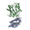 3qn1S S: Starting model for refinement |
|---|---|
| Similar structure data |


-Links
 PDBj
PDBj- Assembly
| Deposited unit | 
| ||||||||
|---|---|---|---|---|---|---|---|---|---|
| 1 |
| ||||||||
| Unit cell |
|
-Components
| #1: Protein | Mass: 20659.332 Da / Num. of mol.: 1 / Mutation: K59R V81I F108A F159L Source method: isolated from a genetically manipulated source Source: (gene. exp.) Arabidopsis thaliana (thale cress) / Gene: PYR1, ABIP6, RCAR11, At4g17870, T6K21.50 / Plasmid: pQE30 / Production host:  Escherichia coli (E. coli) / Strain (production host): BL21 / References: UniProt: O49686 Escherichia coli (E. coli) / Strain (production host): BL21 / References: UniProt: O49686 | ||
|---|---|---|---|
| #2: Protein | Mass: 36582.891 Da / Num. of mol.: 1 Source method: isolated from a genetically manipulated source Source: (gene. exp.) Arabidopsis thaliana (thale cress) / Gene: HAB1, P2C-HA, At1g72770, F28P22.4 / Plasmid: pET11M / Production host: Escherichia coli (E. coli) / Strain (production host): BL21(DE3)References: UniProt: Q9CAJ0, protein-serine/threonine phosphatase | ||
| #3: Chemical | ChemComp-3UZ / (  Mass: 411.878 Da / Num. of mol.: 1 / Source method: obtained synthetically / Formula: C23H22ClNO4 Mass: 411.878 Da / Num. of mol.: 1 / Source method: obtained synthetically / Formula: C23H22ClNO4 | ||
| #4: Chemical |   Mass: 24.305 Da / Num. of mol.: 3 / Source method: obtained synthetically / Formula: Mg Mass: 24.305 Da / Num. of mol.: 3 / Source method: obtained synthetically / Formula: Mg#5: Water | ChemComp-HOH / | Water Mass: 18.015 Da / Num. of mol.: 192 / Source method: isolated from a natural source / Formula: H2O Mass: 18.015 Da / Num. of mol.: 192 / Source method: isolated from a natural source / Formula: H2O |
-Experimental details
-Experiment
| Experiment | Method: X-RAY DIFFRACTION / Number of used crystals: 1 |
|---|
- Sample preparation
| Crystal | Density Matthews: 2.45 Å3/Da / Density % sol: 49.71 % |
|---|---|
| Crystal grow | Temperature: 292 K / Method: vapor diffusion, hanging drop / pH: 7 Details: 22.5% PEG 2000 mono methyl ether 150 mM Sodium malonate |
-Data collection
| Diffraction | Mean temperature: 100 K |
|---|---|
| Diffraction source | Source: ROTATING ANODE / Type: RIGAKU MICROMAX-007 / Wavelength: 1.54 Å |
| Detector | Type: RIGAKU RAXIS IV++ / Detector: IMAGE PLATE / Date: Nov 15, 2013 |
| Radiation | Protocol: SINGLE WAVELENGTH / Monochromatic (M) / Laue (L): M / Scattering type: x-ray |
| Radiation wavelength | Wavelength: 1.54 Å / Relative weight: 1 |
| Reflection | Resolution: 2.25→50 Å / Num. obs: 25330 / % possible obs: 98.5 % / Redundancy: 6.6 % / Rmerge(I) obs: 0.087 / Net I/σ(I): 23.6 |
| Reflection shell | Resolution: 2.25→2.31 Å / Redundancy: 6 % / Rmerge(I) obs: 0.763 / Mean I/σ(I) obs: 2.1 / % possible all: 96.4 |
- Processing
| Software |
| ||||||||||||||||||||||||||||||||||||||||||||||||||||||||||||||||||||||||||||||||||||||||||||||||||
|---|---|---|---|---|---|---|---|---|---|---|---|---|---|---|---|---|---|---|---|---|---|---|---|---|---|---|---|---|---|---|---|---|---|---|---|---|---|---|---|---|---|---|---|---|---|---|---|---|---|---|---|---|---|---|---|---|---|---|---|---|---|---|---|---|---|---|---|---|---|---|---|---|---|---|---|---|---|---|---|---|---|---|---|---|---|---|---|---|---|---|---|---|---|---|---|---|---|---|---|
| Refinement | Method to determine structure: MOLECULAR REPLACEMENT Starting model: PDB ID 3QN1 Resolution: 2.251→36.191 Å / SU ML: 0.26 / Cross valid method: FREE R-VALUE / σ(F): 0 / Phase error: 25.34 / Stereochemistry target values: ML
| ||||||||||||||||||||||||||||||||||||||||||||||||||||||||||||||||||||||||||||||||||||||||||||||||||
| Solvent computation | Shrinkage radii: 0.9 Å / VDW probe radii: 1.11 Å / Solvent model: FLAT BULK SOLVENT MODEL | ||||||||||||||||||||||||||||||||||||||||||||||||||||||||||||||||||||||||||||||||||||||||||||||||||
| Refinement step | Cycle: LAST / Resolution: 2.251→36.191 Å
| ||||||||||||||||||||||||||||||||||||||||||||||||||||||||||||||||||||||||||||||||||||||||||||||||||
| Refine LS restraints |
| ||||||||||||||||||||||||||||||||||||||||||||||||||||||||||||||||||||||||||||||||||||||||||||||||||
| LS refinement shell |
| ||||||||||||||||||||||||||||||||||||||||||||||||||||||||||||||||||||||||||||||||||||||||||||||||||
| Refinement TLS params. | Method: refined / Refine-ID: X-RAY DIFFRACTION
| ||||||||||||||||||||||||||||||||||||||||||||||||||||||||||||||||||||||||||||||||||||||||||||||||||
| Refinement TLS group |
|
