 Movie
Movie Controller


+ Open data

- Basic information
| Entry | Database: PDB / ID: 3kdj | ||||||
|---|---|---|---|---|---|---|---|
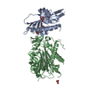
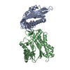
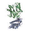
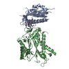
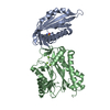
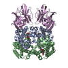
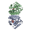
| Title | Complex structure of (+)-ABA-bound PYL1 and ABI1 | ||||||
 Components Components |
| ||||||
 Keywords Keywords | HYDROLASE/Hormone Receptor / ABA / PYL1 /  ABI1 / Abscisic acid signaling pathway / Cell membrane / Hydrolase / Magnesium / Manganese / Metal-binding / Nucleus / Protein phosphatase / HYDROLASE-Hormone Receptor complex ABI1 / Abscisic acid signaling pathway / Cell membrane / Hydrolase / Magnesium / Manganese / Metal-binding / Nucleus / Protein phosphatase / HYDROLASE-Hormone Receptor complex | ||||||
| Function / homology |  Function and homology information Function and homology informationregulation of abscisic acid-activated signaling pathway / negative regulation of abscisic acid-activated signaling pathway / regulation of protein serine/threonine phosphatase activity / protein phosphatase inhibitor complex / regulation of stomatal movement / response to abscisic acid / abscisic acid binding / abscisic acid-activated signaling pathway / protein phosphatase inhibitor activity / plastid ...regulation of abscisic acid-activated signaling pathway / negative regulation of abscisic acid-activated signaling pathway / regulation of protein serine/threonine phosphatase activity / protein phosphatase inhibitor complex / regulation of stomatal movement / response to abscisic acid / abscisic acid binding / abscisic acid-activated signaling pathway / protein phosphatase inhibitor activity / plastid / myosin phosphatase activity / protein serine/threonine phosphatase activity / protein-serine/threonine phosphatase / phosphatase activity / phosphoprotein phosphatase activity / response to cold / protein dephosphorylation / defense response / kinase binding / signaling receptor activity / response to heat / protein kinase binding / protein homodimerization activity / identical protein binding / metal ion binding / nucleus / plasma membrane / cytoplasmSimilarity search - Function | ||||||
| Biological species |  Arabidopsis thaliana (thale cress) Arabidopsis thaliana (thale cress) | ||||||
| Method | X-RAY DIFFRACTION / SYNCHROTRON / MOLECULAR REPLACEMENT / Resolution: 1.878 Å | ||||||
 Authors Authors | Yin, P. / Fan, H. / Hao, Q. / Yuan, X. / Yan, N. | ||||||
 Citation Citation | Journal: Nat.Struct.Mol.Biol. / Year: 2009 Title: Structural insights into the mechanism of abscisic acid signaling by PYL proteins Authors: Yin, P. / Fan, H. / Hao, Q. / Yuan, X. / Wu, D. / Pang, Y. / Yan, C. / Li, W. / Wang, J. / Yan, N. | ||||||
| History |
|
- Structure visualization
| Structure viewer | Molecule: MolmilJmol/JSmol |
|---|
- Downloads & links
-Download
| PDBx/mmCIF format | 3kdj.cif.gz | 199.5 KB | Display | PDBx/mmCIF format |
|---|---|---|---|---|
| PDB format | pdb3kdj.ent.gz | 157.2 KB | Display | PDB format |
| PDBx/mmJSON format | 3kdj.json.gz | Tree view | PDBx/mmJSON format | |
| Others |  Other downloads Other downloads |
-Validation report
| Arichive directory | https://data.pdbj.org/pub/pdb/validation_reports/kd/3kdjftp://data.pdbj.org/pub/pdb/validation_reports/kd/3kdj | HTTPS FTP |
|---|
-Related structure data
| Related structure data |  3kdhSC  3kdiC 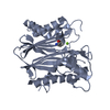 2p8eS S: Starting model for refinement C: citing same article ( |
|---|---|
| Similar structure data |


-Links
 PDBj
PDBj- Assembly
| Deposited unit | 
| ||||||||
|---|---|---|---|---|---|---|---|---|---|
| 1 |
| ||||||||
| Unit cell |
|
-Components
| #1: Protein | Mass: 23327.898 Da / Num. of mol.: 1 / Fragment: residues 20-221 Source method: isolated from a genetically manipulated source Source: (gene. exp.) Arabidopsis thaliana (thale cress) / Gene: At5g46790 / Plasmid: pET-15b / Production host:  Escherichia coli (E. coli) / Strain (production host): BL21(DE3) / References: UniProt: Q8VZS8 Escherichia coli (E. coli) / Strain (production host): BL21(DE3) / References: UniProt: Q8VZS8 |
|---|---|
| #2: Protein | Mass: 34792.836 Da / Num. of mol.: 1 / Fragment: residues 119-434 / Mutation: C208S, D378G Source method: isolated from a genetically manipulated source Source: (gene. exp.) Arabidopsis thaliana (thale cress) / Gene: ABI1, At4g26080, F20B18.190 / Plasmid: pET-15b / Production host: Escherichia coli (E. coli) / Strain (production host): BL21(DE3)References: UniProt: P49597, protein-serine/threonine phosphatase |
| #3: Chemical | ChemComp-A8S / (Abscisic acid  Mass: 264.317 Da / Num. of mol.: 1 / Source method: obtained synthetically / Formula: C15H20O4 / Comment: hormone*YM Mass: 264.317 Da / Num. of mol.: 1 / Source method: obtained synthetically / Formula: C15H20O4 / Comment: hormone*YM |
| #4: Chemical | ChemComp-MN /   Mass: 54.938 Da / Num. of mol.: 1 / Source method: obtained synthetically / Formula: Mn Mass: 54.938 Da / Num. of mol.: 1 / Source method: obtained synthetically / Formula: Mn |
| #5: Water | ChemComp-HOH / Water Mass: 18.015 Da / Num. of mol.: 170 / Source method: isolated from a natural source / Formula: H2O Mass: 18.015 Da / Num. of mol.: 170 / Source method: isolated from a natural source / Formula: H2O |
-Experimental details
-Experiment
| Experiment | Method: X-RAY DIFFRACTION / Number of used crystals: 1 |
|---|
- Sample preparation
| Crystal | Density Matthews: 2.55 Å3/Da / Density % sol: 51.69 % |
|---|---|
| Crystal grow | Temperature: 291 K / Method: vapor diffusion, hanging drop / pH: 5.5 Details: 0.1M citric acid pH 5.5, 0.01M GSH/GSSG, 10% PEG 4000, VAPOR DIFFUSION, HANGING DROP, temperature 291K |
-Data collection
| Diffraction | Mean temperature: 100 K |
|---|---|
| Diffraction source | Source: SYNCHROTRON / Site: SPring-8  / Beamline: BL41XU / Wavelength: 1 Å / Beamline: BL41XU / Wavelength: 1 Å |
| Detector | Type: MARMOSAIC 225 mm CCD / Detector: CCD / Date: Oct 16, 2009 |
| Radiation | Protocol: SINGLE WAVELENGTH / Monochromatic (M) / Laue (L): M / Scattering type: x-ray |
| Radiation wavelength | Wavelength: 1 Å / Relative weight: 1 |
| Reflection | Resolution: 1.878→34.15 Å / Num. obs: 44478 / % possible obs: 90.4 % / Redundancy: 4.2 % / Biso Wilson estimate: 34.99 Å2 / Rmerge(I) obs: 0.053 / Net I/σ(I): 24.75 |
| Reflection shell | Resolution: 1.878→1.95 Å / Redundancy: 3.3 % / Rmerge(I) obs: 0.594 / Mean I/σ(I) obs: 1.87 / Num. unique all: 2581 / % possible all: 53.2 |
- Processing
| Software |
| |||||||||||||||||||||||||||||||||||||||||||||||||||||||||||||||||||||||||||||||||||||||||||||||||||||||||||||||||||||||
|---|---|---|---|---|---|---|---|---|---|---|---|---|---|---|---|---|---|---|---|---|---|---|---|---|---|---|---|---|---|---|---|---|---|---|---|---|---|---|---|---|---|---|---|---|---|---|---|---|---|---|---|---|---|---|---|---|---|---|---|---|---|---|---|---|---|---|---|---|---|---|---|---|---|---|---|---|---|---|---|---|---|---|---|---|---|---|---|---|---|---|---|---|---|---|---|---|---|---|---|---|---|---|---|---|---|---|---|---|---|---|---|---|---|---|---|---|---|---|---|---|
| Refinement | Method to determine structure: MOLECULAR REPLACEMENT Starting model: PDB ENTRIES: 3KDH and 2P8E Resolution: 1.878→34.15 Å / Occupancy max: 1 / Occupancy min: 0.33 / FOM work R set: 0.718 / SU ML: 0.28 / Isotropic thermal model: TLS / σ(F): 1.34 / Phase error: 33.13 / Stereochemistry target values: ML
| |||||||||||||||||||||||||||||||||||||||||||||||||||||||||||||||||||||||||||||||||||||||||||||||||||||||||||||||||||||||
| Solvent computation | Shrinkage radii: 0.9 Å / VDW probe radii: 1.11 Å / Solvent model: FLAT BULK SOLVENT MODEL / Bsol: 59.253 Å2 / ksol: 0.345 e/Å3 | |||||||||||||||||||||||||||||||||||||||||||||||||||||||||||||||||||||||||||||||||||||||||||||||||||||||||||||||||||||||
| Displacement parameters | Biso max: 260.35 Å2 / Biso mean: 60.477 Å2 / Biso min: 30.46 Å2
| |||||||||||||||||||||||||||||||||||||||||||||||||||||||||||||||||||||||||||||||||||||||||||||||||||||||||||||||||||||||
| Refinement step | Cycle: LAST / Resolution: 1.878→34.15 Å
| |||||||||||||||||||||||||||||||||||||||||||||||||||||||||||||||||||||||||||||||||||||||||||||||||||||||||||||||||||||||
| Refine LS restraints |
| |||||||||||||||||||||||||||||||||||||||||||||||||||||||||||||||||||||||||||||||||||||||||||||||||||||||||||||||||||||||
| LS refinement shell |
| |||||||||||||||||||||||||||||||||||||||||||||||||||||||||||||||||||||||||||||||||||||||||||||||||||||||||||||||||||||||
| Refinement TLS params. | Method: refined / Refine-ID: X-RAY DIFFRACTION
| |||||||||||||||||||||||||||||||||||||||||||||||||||||||||||||||||||||||||||||||||||||||||||||||||||||||||||||||||||||||
| Refinement TLS group |
|
