 Movie
Movie Controller

[English] 日本語
 Yorodumi
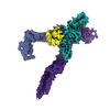
- PDB-8c7m: Interleukin 12 receptor subunit beta-1 Fn domains in complex with... -
+ Open data

- Basic information
| Entry | Database: PDB / ID: 8c7m | |||||||||
|---|---|---|---|---|---|---|---|---|---|---|
| Title | Interleukin 12 receptor subunit beta-1 Fn domains in complex with antagonistic FAb fragment. | |||||||||
 Components Components |
| |||||||||
 Keywords Keywords |  CYTOKINE / receptor / antagonist / antibody CYTOKINE / receptor / antagonist / antibody | |||||||||
| Function / homology |  Function and homology information Function and homology informationinterleukin-23-mediated signaling pathway / positive regulation of T-helper 1 type immune response / positive regulation of memory T cell differentiation / interleukin-12 receptor complex / interleukin-23 receptor complex / Interleukin-23 signaling / positive regulation of T-helper 17 type immune response / interleukin-12-mediated signaling pathway / Interleukin-12 signaling / cytokine receptor activity ...interleukin-23-mediated signaling pathway / positive regulation of T-helper 1 type immune response / positive regulation of memory T cell differentiation / interleukin-12 receptor complex / interleukin-23 receptor complex / Interleukin-23 signaling / positive regulation of T-helper 17 type immune response / interleukin-12-mediated signaling pathway / Interleukin-12 signaling / cytokine receptor activity / positive regulation of activated T cell proliferation / cytokine binding / positive regulation of T-helper 17 cell lineage commitment / positive regulation of defense response to virus by host / cytokine-mediated signaling pathway / cellular response to type II interferon / positive regulation of T cell mediated cytotoxicity / positive regulation of type II interferon production / receptor complex / external side of plasma membrane / signal transduction / plasma membraneSimilarity search - Function | |||||||||
| Biological species |  Homo sapiens (human) Homo sapiens (human) Homo sapiens x Mus musculus hybrid cell line (mammal) Homo sapiens x Mus musculus hybrid cell line (mammal) | |||||||||
| Method | X-RAY DIFFRACTION / SYNCHROTRON / MOLECULAR REPLACEMENT / Resolution: 2.56 Å | |||||||||
 Authors Authors | Bloch, Y. / Savvides, S.N. | |||||||||
| Funding support |  Belgium, 2items Belgium, 2items
| |||||||||
 Citation Citation | Journal: Nat Struct Mol Biol / Year: 2024 Title: Structures of complete extracellular receptor assemblies mediated by IL-12 and IL-23. Authors: Yehudi Bloch / Jan Felix / Romain Merceron / Mathias Provost / Royan Alipour Symakani / Robin De Backer / Elisabeth Lambert / Ahmad R Mehdipour / Savvas N Savvides /    Abstract: Cell-surface receptor complexes mediated by pro-inflammatory interleukin (IL)-12 and IL-23, both validated therapeutic targets, are incompletely understood due to the lack of structural insights into ...Cell-surface receptor complexes mediated by pro-inflammatory interleukin (IL)-12 and IL-23, both validated therapeutic targets, are incompletely understood due to the lack of structural insights into their complete extracellular assemblies. Furthermore, there is a paucity of structural details describing the IL-12-receptor interaction interfaces, in contrast to IL-23-receptor complexes. Here we report structures of fully assembled mouse IL-12/human IL-23-receptor complexes comprising the complete extracellular segments of the cognate receptors determined by electron cryo-microscopy. The structures reveal key commonalities but also surprisingly diverse features. Most notably, whereas IL-12 and IL-23 both utilize a conspicuously presented aromatic residue on their α-subunit as a hotspot to interact with the N-terminal Ig domain of their high-affinity receptors, only IL-12 juxtaposes receptor domains proximal to the cell membrane. Collectively, our findings will help to complete our understanding of cytokine-mediated assemblies of tall cytokine receptors and will enable a cytokine-specific interrogation of IL-12/IL-23 signaling in physiology and disease. | |||||||||
| History |
|
- Structure visualization
| Structure viewer | Molecule: MolmilJmol/JSmol |
|---|
- Downloads & links
-Download
| PDBx/mmCIF format | 8c7m.cif.gz | 563.6 KB | Display | PDBx/mmCIF format |
|---|---|---|---|---|
| PDB format | pdb8c7m.ent.gz | 468.4 KB | Display | PDB format |
| PDBx/mmJSON format | 8c7m.json.gz | Tree view | PDBx/mmJSON format | |
| Others |  Other downloads Other downloads |
-Validation report
| Arichive directory | https://data.pdbj.org/pub/pdb/validation_reports/c7/8c7mftp://data.pdbj.org/pub/pdb/validation_reports/c7/8c7m | HTTPS FTP |
|---|
-Related structure data
| Related structure data |  8cr5C  8cr6C  8cr8C  8odxC  8odzC  8oe0C  8oe4C  8pb1C  8ppmC C: citing same article ( |
|---|---|
| Similar structure data |


-Links
 PDBj
PDBj


- Assembly
| Deposited unit | 
| ||||||||
|---|---|---|---|---|---|---|---|---|---|
| 1 |
| ||||||||
| 2 |
| ||||||||
| Unit cell |
|
-Components
-Antibody , 2 types, 4 molecules CHDL
| #2: Antibody | Mass: 24786.586 Da / Num. of mol.: 2 Source method: isolated from a genetically manipulated source Details: [225-231] encode cloning scar and 6His tag Source: (gene. exp.) Homo sapiens x Mus musculus hybrid cell line (mammal)Plasmid: pHLsec / Cell line (production host): HEK293S / Production host: Homo sapiens (human)#3: Antibody | Mass: 23239.732 Da / Num. of mol.: 2 Source method: isolated from a genetically manipulated source Source: (gene. exp.) Homo sapiens x Mus musculus hybrid cell line (mammal)Plasmid: pHLsec / Cell line (production host): HEK293S MGAT1-/- / Production host: Homo sapiens (human) |
|---|
-Protein / Sugars , 2 types, 7 molecules AB
| #1: Protein | / IL-12 receptor subunit beta-1 / IL-12R subunit beta-1 / IL-12R-beta-1 / IL-12RB1 / IL-12 receptor ...IL-12 receptor subunit beta-1 / IL-12R subunit beta-1 / IL-12R-beta-1 / IL-12RB1 / IL-12 receptor beta component Mass: 34256.363 Da / Num. of mol.: 2 Source method: isolated from a genetically manipulated source Details: [310-314] encode a protease site / Source: (gene. exp.) Homo sapiens (human) / Gene: IL12RB1, IL12R, IL12RB / Plasmid: pHLsec / Cell line (production host): HEK293S MGAT1-/- / Production host: Homo sapiens (human) / References: UniProt: P42701#4: Sugar | ChemComp-NAG / N-Acetylglucosamine Type: D-saccharide, beta linking / Mass: 221.208 Da / Num. of mol.: 5 Type: D-saccharide, beta linking / Mass: 221.208 Da / Num. of mol.: 5Source method: isolated from a genetically manipulated source Formula: C8H15NO6 |
|---|
-Non-polymers , 2 types, 26 molecules 

| #5: Chemical | Chloride Mass: 35.453 Da / Num. of mol.: 2 / Source method: obtained synthetically / Formula: Cl Mass: 35.453 Da / Num. of mol.: 2 / Source method: obtained synthetically / Formula: Cl#6: Water | ChemComp-HOH / | WaterMass: 18.015 Da / Num. of mol.: 24 / Source method: isolated from a natural source / Formula: H2O |
|---|
-Details
| Has ligand of interest | N |
|---|
-Experimental details
-Experiment
| Experiment | Method: X-RAY DIFFRACTION / Number of used crystals: 1 |
|---|
- Sample preparation
| Crystal | Density Matthews: 3.44 Å3/Da / Density % sol: 64.26 % |
|---|---|
| Crystal grow | Temperature: 287 K / Method: vapor diffusion, sitting drop Details: Morpheus2 condition A3: 90 mM LiNaKSO4, 100 mM Citrate BisTRIS propane pH 5.4, 8.57% PEG 8000, 17.14% 1,5 Pentanediol |
-Data collection
| Diffraction | Mean temperature: 100 K / Serial crystal experiment: N | ||||||||||||||||||||||||||||||||||||||||||||||||||||||||||||||||||||||
|---|---|---|---|---|---|---|---|---|---|---|---|---|---|---|---|---|---|---|---|---|---|---|---|---|---|---|---|---|---|---|---|---|---|---|---|---|---|---|---|---|---|---|---|---|---|---|---|---|---|---|---|---|---|---|---|---|---|---|---|---|---|---|---|---|---|---|---|---|---|---|---|
| Diffraction source | Source: SYNCHROTRON / Site: PETRA III, EMBL c/o DESY / Beamline: P14 (MX2) / Wavelength: 0.9763 Å | ||||||||||||||||||||||||||||||||||||||||||||||||||||||||||||||||||||||
| Detector | Type: DECTRIS EIGER X 16M / Detector: PIXEL / Date: Aug 16, 2021 | ||||||||||||||||||||||||||||||||||||||||||||||||||||||||||||||||||||||
| Radiation | Protocol: SINGLE WAVELENGTH / Monochromatic (M) / Laue (L): M / Scattering type: x-ray | ||||||||||||||||||||||||||||||||||||||||||||||||||||||||||||||||||||||
| Radiation wavelength | Wavelength: 0.9763 Å / Relative weight: 1 | ||||||||||||||||||||||||||||||||||||||||||||||||||||||||||||||||||||||
| Reflection | Resolution: 2.56→140.18 Å / Num. obs: 73952 / % possible obs: 99.8 % / Redundancy: 13.6 % / Biso Wilson estimate: 76.27 Å2 / CC1/2: 0.999 / Rrim(I) all: 0.104 / Net I/σ(I): 19.16 | ||||||||||||||||||||||||||||||||||||||||||||||||||||||||||||||||||||||
| Reflection shell | Diffraction-ID: 1
|
- Processing
| Software |
| |||||||||||||||||||||||||||||||||||||||||||||||||||||||||||||||||||||||||||||||||||||||||||||||||||||||||||||||||||||||||||||||||||||||||||||||||||||||||||||||||||||||||||||||||||||||||||||||||||||||||||||||||||||||||||||||||||||||||||||||||||||||||||||||||||||||||||||||||||||||||||||||||||||||||||||||||||||||||||||||||||||||||||||||||||||||||||||||||||||||||||||||||||||||
|---|---|---|---|---|---|---|---|---|---|---|---|---|---|---|---|---|---|---|---|---|---|---|---|---|---|---|---|---|---|---|---|---|---|---|---|---|---|---|---|---|---|---|---|---|---|---|---|---|---|---|---|---|---|---|---|---|---|---|---|---|---|---|---|---|---|---|---|---|---|---|---|---|---|---|---|---|---|---|---|---|---|---|---|---|---|---|---|---|---|---|---|---|---|---|---|---|---|---|---|---|---|---|---|---|---|---|---|---|---|---|---|---|---|---|---|---|---|---|---|---|---|---|---|---|---|---|---|---|---|---|---|---|---|---|---|---|---|---|---|---|---|---|---|---|---|---|---|---|---|---|---|---|---|---|---|---|---|---|---|---|---|---|---|---|---|---|---|---|---|---|---|---|---|---|---|---|---|---|---|---|---|---|---|---|---|---|---|---|---|---|---|---|---|---|---|---|---|---|---|---|---|---|---|---|---|---|---|---|---|---|---|---|---|---|---|---|---|---|---|---|---|---|---|---|---|---|---|---|---|---|---|---|---|---|---|---|---|---|---|---|---|---|---|---|---|---|---|---|---|---|---|---|---|---|---|---|---|---|---|---|---|---|---|---|---|---|---|---|---|---|---|---|---|---|---|---|---|---|---|---|---|---|---|---|---|---|---|---|---|---|---|---|---|---|---|---|---|---|---|---|---|---|---|---|---|---|---|---|---|---|---|---|---|---|---|---|---|---|---|---|---|---|---|---|---|---|---|---|---|---|---|---|---|---|---|---|---|---|---|---|---|---|---|---|---|---|---|---|---|---|---|---|---|---|---|---|---|---|---|---|---|---|---|---|---|---|---|---|---|---|---|---|---|---|---|---|
| Refinement | Method to determine structure: MOLECULAR REPLACEMENT / Resolution: 2.56→70.09 Å / SU ML: 0.38 / Cross valid method: FREE R-VALUE / σ(F): 1.34 / Phase error: 25.35 / Stereochemistry target values: ML
| |||||||||||||||||||||||||||||||||||||||||||||||||||||||||||||||||||||||||||||||||||||||||||||||||||||||||||||||||||||||||||||||||||||||||||||||||||||||||||||||||||||||||||||||||||||||||||||||||||||||||||||||||||||||||||||||||||||||||||||||||||||||||||||||||||||||||||||||||||||||||||||||||||||||||||||||||||||||||||||||||||||||||||||||||||||||||||||||||||||||||||||||||||||||
| Solvent computation | Shrinkage radii: 0.9 Å / VDW probe radii: 1.11 Å / Solvent model: FLAT BULK SOLVENT MODEL | |||||||||||||||||||||||||||||||||||||||||||||||||||||||||||||||||||||||||||||||||||||||||||||||||||||||||||||||||||||||||||||||||||||||||||||||||||||||||||||||||||||||||||||||||||||||||||||||||||||||||||||||||||||||||||||||||||||||||||||||||||||||||||||||||||||||||||||||||||||||||||||||||||||||||||||||||||||||||||||||||||||||||||||||||||||||||||||||||||||||||||||||||||||||
| Refinement step | Cycle: LAST / Resolution: 2.56→70.09 Å
| |||||||||||||||||||||||||||||||||||||||||||||||||||||||||||||||||||||||||||||||||||||||||||||||||||||||||||||||||||||||||||||||||||||||||||||||||||||||||||||||||||||||||||||||||||||||||||||||||||||||||||||||||||||||||||||||||||||||||||||||||||||||||||||||||||||||||||||||||||||||||||||||||||||||||||||||||||||||||||||||||||||||||||||||||||||||||||||||||||||||||||||||||||||||
| Refine LS restraints |
| |||||||||||||||||||||||||||||||||||||||||||||||||||||||||||||||||||||||||||||||||||||||||||||||||||||||||||||||||||||||||||||||||||||||||||||||||||||||||||||||||||||||||||||||||||||||||||||||||||||||||||||||||||||||||||||||||||||||||||||||||||||||||||||||||||||||||||||||||||||||||||||||||||||||||||||||||||||||||||||||||||||||||||||||||||||||||||||||||||||||||||||||||||||||
| LS refinement shell |
| |||||||||||||||||||||||||||||||||||||||||||||||||||||||||||||||||||||||||||||||||||||||||||||||||||||||||||||||||||||||||||||||||||||||||||||||||||||||||||||||||||||||||||||||||||||||||||||||||||||||||||||||||||||||||||||||||||||||||||||||||||||||||||||||||||||||||||||||||||||||||||||||||||||||||||||||||||||||||||||||||||||||||||||||||||||||||||||||||||||||||||||||||||||||
| Refinement TLS params. | Method: refined / Refine-ID: X-RAY DIFFRACTION
| |||||||||||||||||||||||||||||||||||||||||||||||||||||||||||||||||||||||||||||||||||||||||||||||||||||||||||||||||||||||||||||||||||||||||||||||||||||||||||||||||||||||||||||||||||||||||||||||||||||||||||||||||||||||||||||||||||||||||||||||||||||||||||||||||||||||||||||||||||||||||||||||||||||||||||||||||||||||||||||||||||||||||||||||||||||||||||||||||||||||||||||||||||||||
| Refinement TLS group |
|
