 Movie
Movie Controller


+ Open data

- Basic information
| Entry | Database: PDB / ID: 7z11 | ||||||
|---|---|---|---|---|---|---|---|
| Title | Structure of substrate bound DRG1 (AFG2) | ||||||
 Components Components |
| ||||||
 Keywords Keywords |  CHAPERONE / Ribosome biogenesis / AAA-ATPases / substrate recognition / single particle cryo-EM CHAPERONE / Ribosome biogenesis / AAA-ATPases / substrate recognition / single particle cryo-EM | ||||||
| Function / homology |  Function and homology information Function and homology informationprotein hexamerization / non-chaperonin molecular chaperone ATPase / preribosome, large subunit precursor / ribosomal large subunit biogenesis / response to xenobiotic stimulus / ATP hydrolysis activity / ATP binding / cytoplasmSimilarity search - Function | ||||||
| Biological species |  Saccharomyces cerevisiae S288C (yeast) Saccharomyces cerevisiae S288C (yeast) | ||||||
| Method | ELECTRON MICROSCOPY / single particle reconstruction / cryo EM / Resolution: 3.2 Å | ||||||
 Authors Authors | Prattes, M. / Grishkovskaya, I. / Bergler, H. / Haselbach, D. | ||||||
| Funding support |  Austria, 1items Austria, 1items
| ||||||
 Citation Citation | Journal: Nat Struct Mol Biol / Year: 2022 Title: Visualizing maturation factor extraction from the nascent ribosome by the AAA-ATPase Drg1. Authors: Michael Prattes / Irina Grishkovskaya / Victor-Valentin Hodirnau / Christina Hetzmannseder / Gertrude Zisser / Carolin Sailer / Vasileios Kargas / Mathias Loibl / Magdalena Gerhalter / Lisa ...Authors: Michael Prattes / Irina Grishkovskaya / Victor-Valentin Hodirnau / Christina Hetzmannseder / Gertrude Zisser / Carolin Sailer / Vasileios Kargas / Mathias Loibl / Magdalena Gerhalter / Lisa Kofler / Alan J Warren / Florian Stengel / David Haselbach / Helmut Bergler /   Abstract: The AAA-ATPase Drg1 is a key factor in eukaryotic ribosome biogenesis that initiates cytoplasmic maturation of the large ribosomal subunit. Drg1 releases the shuttling maturation factor Rlp24 from ...The AAA-ATPase Drg1 is a key factor in eukaryotic ribosome biogenesis that initiates cytoplasmic maturation of the large ribosomal subunit. Drg1 releases the shuttling maturation factor Rlp24 from pre-60S particles shortly after nuclear export, a strict requirement for downstream maturation. The molecular mechanism of release remained elusive. Here, we report a series of cryo-EM structures that captured the extraction of Rlp24 from pre-60S particles by Saccharomyces cerevisiae Drg1. These structures reveal that Arx1 and the eukaryote-specific rRNA expansion segment ES27 form a joint docking platform that positions Drg1 for efficient extraction of Rlp24 from the pre-ribosome. The tips of the Drg1 N domains thereby guide the Rlp24 C terminus into the central pore of the Drg1 hexamer, enabling extraction by a hand-over-hand translocation mechanism. Our results uncover substrate recognition and processing by Drg1 step by step and provide a comprehensive mechanistic picture of the conserved modus operandi of AAA-ATPases. | ||||||
| History |
|
- Structure visualization
| Structure viewer | Molecule: MolmilJmol/JSmol |
|---|
- Downloads & links
-Download
| PDBx/mmCIF format | 7z11.cif.gz | 718.1 KB | Display | PDBx/mmCIF format |
|---|---|---|---|---|
| PDB format | pdb7z11.ent.gz | 608.1 KB | Display | PDB format |
| PDBx/mmJSON format | 7z11.json.gz | Tree view | PDBx/mmJSON format | |
| Others |  Other downloads Other downloads |
-Validation report
| Arichive directory | https://data.pdbj.org/pub/pdb/validation_reports/z1/7z11ftp://data.pdbj.org/pub/pdb/validation_reports/z1/7z11 | HTTPS FTP |
|---|
-Related structure data
| Related structure data |  14437MC 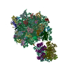 7z34C M: map data used to model this data C: citing same article ( |
|---|---|
| Similar structure data |

-Links
 PDBj
PDBj


- Assembly
| Deposited unit | 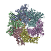
|
|---|---|
| 1 |
|
-Components
| #1: Protein | Mass: 84850.719 Da / Num. of mol.: 6 Source method: isolated from a genetically manipulated source Source: (gene. exp.) Saccharomyces cerevisiae S288C (yeast) / Strain: ATCC 204508 / S288c / Gene: AFG2, DRG1, YLR397C, L8084.16 / Production host: Saccharomyces cerevisiae BY4743 (yeast)References: UniProt: P32794, non-chaperonin molecular chaperone ATPase#2: Protein/peptide | | Mass: 1720.111 Da / Num. of mol.: 1 Source method: isolated from a genetically manipulated source Source: (gene. exp.) Saccharomyces cerevisiae S288C (yeast) / Production host: Saccharomyces cerevisiae BY4743 (yeast)#3: Chemical | ChemComp-AGS /   Mass: 523.247 Da / Num. of mol.: 11 / Source method: obtained synthetically / Formula: C10H16N5O12P3S / Feature type: SUBJECT OF INVESTIGATION / Comment: ATP-gamma-S, energy-carrying molecule analogue*YM Mass: 523.247 Da / Num. of mol.: 11 / Source method: obtained synthetically / Formula: C10H16N5O12P3S / Feature type: SUBJECT OF INVESTIGATION / Comment: ATP-gamma-S, energy-carrying molecule analogue*YMHas ligand of interest | Y | |
|---|
-Experimental details
-Experiment
| Experiment | Method: ELECTRON MICROSCOPY |
|---|---|
| EM experiment | Aggregation state: PARTICLE / 3D reconstruction method: single particle reconstruction |
- Sample preparation
| Component | Name: AFG2 hexamer / Type: COMPLEX / Entity ID: #1-#2 / Source: RECOMBINANT |
|---|---|
| Source (natural) | Organism: Saccharomyces cerevisiae S288C (yeast) |
| Source (recombinant) | Organism: Saccharomyces cerevisiae BY4743 (yeast) |
| Buffer solution | pH: 7.6 |
| Specimen | Embedding applied: NO / Shadowing applied: NO / Staining applied: NO / Vitrification applied: YES |
| Specimen support | Grid material: COPPER / Grid mesh size: 200 divisions/in. / Grid type: Quantifoil R1.2/1.3 |
| Vitrification | Instrument: FEI VITROBOT MARK IV / Cryogen name: ETHANE / Humidity: 100 % / Chamber temperature: 277.15 K |
- Electron microscopy imaging
| Experimental equipment |  Model: Titan Krios / Image courtesy: FEI Company |
|---|---|
| Microscopy | Model: FEI TITAN KRIOS |
| Electron gun | Electron source: FIELD EMISSION GUN / Accelerating voltage: 300 kV / Illumination mode: FLOOD BEAM |
| Electron lens | Mode: BRIGHT FIELDBright-field microscopy / Nominal defocus max: 2500 nm / Nominal defocus min: 500 nm / Cs: 2.7 mm / C2 aperture diameter: 50 µm / Alignment procedure: COMA FREE |
| Specimen holder | Cryogen: NITROGEN / Specimen holder model: FEI TITAN KRIOS AUTOGRID HOLDER |
| Image recording | Electron dose: 60 e/Å2 / Film or detector model: GATAN K3 BIOQUANTUM (6k x 4k) |
| EM imaging optics | Energyfilter slit width: 20 eV |
- Processing
| Software | Name: PHENIX / Version: 1.18.2_3874: / Classification: refinement | ||||||||||||||||||||||||||||||||||||
|---|---|---|---|---|---|---|---|---|---|---|---|---|---|---|---|---|---|---|---|---|---|---|---|---|---|---|---|---|---|---|---|---|---|---|---|---|---|
| EM software |
| ||||||||||||||||||||||||||||||||||||
| CTF correction | Type: PHASE FLIPPING AND AMPLITUDE CORRECTION | ||||||||||||||||||||||||||||||||||||
| Particle selection | Num. of particles selected: 3148330 | ||||||||||||||||||||||||||||||||||||
| 3D reconstruction | Resolution: 3.2 Å / Resolution method: FSC 0.143 CUT-OFF / Num. of particles: 114728 / Symmetry type: POINT | ||||||||||||||||||||||||||||||||||||
| Atomic model building | Protocol: AB INITIO MODEL / Space: REAL / Target criteria: correlation coefficient | ||||||||||||||||||||||||||||||||||||
| Refine LS restraints |
|
