 Movie
Movie Controller


+ Open data

- Basic information
| Entry |  | |||||||||
|---|---|---|---|---|---|---|---|---|---|---|
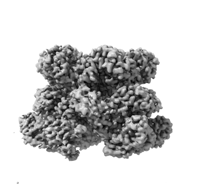
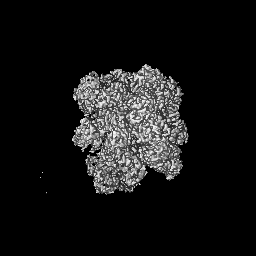
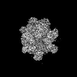
| Title | Structure of substrate bound DRG1 (AFG2) | |||||||||
 Map data Map data | ||||||||||
 Sample Sample |
| |||||||||
| Function / homology |  Function and homology information Function and homology informationprotein hexamerization /  non-chaperonin molecular chaperone ATPase / preribosome, large subunit precursor / ribosomal large subunit biogenesis / response to xenobiotic stimulus / ATP hydrolysis activity / ATP binding / cytoplasm non-chaperonin molecular chaperone ATPase / preribosome, large subunit precursor / ribosomal large subunit biogenesis / response to xenobiotic stimulus / ATP hydrolysis activity / ATP binding / cytoplasmSimilarity search - Function | |||||||||
| Biological species |  Saccharomyces cerevisiae S288C (yeast) Saccharomyces cerevisiae S288C (yeast) | |||||||||
| Method | single particle reconstruction / cryo EM / Resolution: 3.2 Å | |||||||||
 Authors Authors | Prattes M / Grishkovskaya I / Bergler H / Haselbach D | |||||||||
| Funding support |  Austria, 1 items Austria, 1 items
| |||||||||
 Citation Citation | Journal: Nat Struct Mol Biol / Year: 2022 Title: Visualizing maturation factor extraction from the nascent ribosome by the AAA-ATPase Drg1. Authors: Michael Prattes / Irina Grishkovskaya / Victor-Valentin Hodirnau / Christina Hetzmannseder / Gertrude Zisser / Carolin Sailer / Vasileios Kargas / Mathias Loibl / Magdalena Gerhalter / Lisa ...Authors: Michael Prattes / Irina Grishkovskaya / Victor-Valentin Hodirnau / Christina Hetzmannseder / Gertrude Zisser / Carolin Sailer / Vasileios Kargas / Mathias Loibl / Magdalena Gerhalter / Lisa Kofler / Alan J Warren / Florian Stengel / David Haselbach / Helmut Bergler /   Abstract: The AAA-ATPase Drg1 is a key factor in eukaryotic ribosome biogenesis that initiates cytoplasmic maturation of the large ribosomal subunit. Drg1 releases the shuttling maturation factor Rlp24 from ...The AAA-ATPase Drg1 is a key factor in eukaryotic ribosome biogenesis that initiates cytoplasmic maturation of the large ribosomal subunit. Drg1 releases the shuttling maturation factor Rlp24 from pre-60S particles shortly after nuclear export, a strict requirement for downstream maturation. The molecular mechanism of release remained elusive. Here, we report a series of cryo-EM structures that captured the extraction of Rlp24 from pre-60S particles by Saccharomyces cerevisiae Drg1. These structures reveal that Arx1 and the eukaryote-specific rRNA expansion segment ES27 form a joint docking platform that positions Drg1 for efficient extraction of Rlp24 from the pre-ribosome. The tips of the Drg1 N domains thereby guide the Rlp24 C terminus into the central pore of the Drg1 hexamer, enabling extraction by a hand-over-hand translocation mechanism. Our results uncover substrate recognition and processing by Drg1 step by step and provide a comprehensive mechanistic picture of the conserved modus operandi of AAA-ATPases. | |||||||||
| History |
|
- Structure visualization
| Supplemental images |
|---|
- Downloads & links
-EMDB archive
| Map data | emd_14437.map.gz | 54.9 MB | EMDB map data format | |
|---|---|---|---|---|
| Header (meta data) | emd-14437-v30.xmlemd-14437.xml | 14.7 KB 14.7 KB | Display Display | EMDB header |
| FSC (resolution estimation) | emd_14437_fsc.xml | 11.7 KB | Display | FSC data file |
| Images |  emd_14437.png emd_14437.png | 76.7 KB | ||
| Archive directory |  http://ftp.pdbj.org/pub/emdb/structures/EMD-14437ftp://ftp.pdbj.org/pub/emdb/structures/EMD-14437 http://ftp.pdbj.org/pub/emdb/structures/EMD-14437ftp://ftp.pdbj.org/pub/emdb/structures/EMD-14437 | HTTPS FTP |
-Related structure data
| Related structure data | 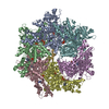 7z11MC 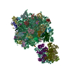 7z34C M: atomic model generated by this map C: citing same article ( |
|---|---|
| Similar structure data |

-Links
| EMDB pages | EMDB (EBI/PDBe) / EMDataResource |
|---|---|
| Related items in Molecule of the Month |


-Map
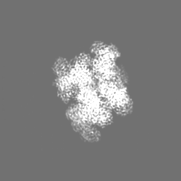
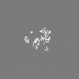
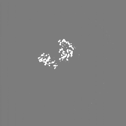
| File | Download / File: emd_14437.map.gz / Format: CCP4 / Size: 64 MB / Type: IMAGE STORED AS FLOATING POINT NUMBER (4 BYTES) | ||||||||||||||||||||||||||||||||||||
|---|---|---|---|---|---|---|---|---|---|---|---|---|---|---|---|---|---|---|---|---|---|---|---|---|---|---|---|---|---|---|---|---|---|---|---|---|---|
| Projections & slices | Image control
Images are generated by Spider. | ||||||||||||||||||||||||||||||||||||
| Voxel size | X=Y=Z: 1.07 Å | ||||||||||||||||||||||||||||||||||||
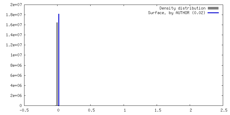
| Density |
| ||||||||||||||||||||||||||||||||||||
| Symmetry | Space group: 1 | ||||||||||||||||||||||||||||||||||||
| Details | EMDB XML:
|
 Z (Sec.)
 Y (Row.)
 X (Col.)


-Supplemental data
- Sample components
-Entire : AFG2 hexamer
| Entire | Name: AFG2 hexamer |
|---|---|
| Components |
|
-Supramolecule #1: AFG2 hexamer
| Supramolecule | Name: AFG2 hexamer / type: complex / Chimera: Yes / ID: 1 / Parent: 0 / Macromolecule list: #1-#2 |
|---|---|
| Source (natural) | Organism: Saccharomyces cerevisiae S288C (yeast) |
| Recombinant expression | Organism: Saccharomyces cerevisiae BY4743 (yeast) |
-Macromolecule #1: ATPase family gene 2 protein
| Macromolecule | Name: ATPase family gene 2 protein / type: protein_or_peptide / ID: 1 / Number of copies: 6 / Enantiomer: LEVO / EC number: non-chaperonin molecular chaperone ATPase |
|---|---|
| Source (natural) | Organism: Saccharomyces cerevisiae S288C (yeast) / Strain: ATCC 204508 / S288c |
| Molecular weight | Theoretical: 84.850719 KDa |
| Recombinant expression | Organism: Saccharomyces cerevisiae BY4743 (yeast) |
| Sequence | String: MAPKSSSSGS KKKSSASSNS ADAKASKFKL PAEFITRPHP SKDHGKETCT AYIHPNVLSS LEINPGSFCT VGKIGENGIL VIARAGDEE VHPVNVITLS TTIRSVGNLI LGDRLELKKA QVQPPYATKV TVGSLQGYNI LECMEEKVIQ KLLDDSGVIM P GMIFQNLK ...String: MAPKSSSSGS KKKSSASSNS ADAKASKFKL PAEFITRPHP SKDHGKETCT AYIHPNVLSS LEINPGSFCT VGKIGENGIL VIARAGDEE VHPVNVITLS TTIRSVGNLI LGDRLELKKA QVQPPYATKV TVGSLQGYNI LECMEEKVIQ KLLDDSGVIM P GMIFQNLK TKAGDESIDV VITDASDDSL PDVSQLDLNM DDMYGGLDNL FYLSPPFIFR KGSTHITFSK ETQANRKYNL PE PLSYAAV GGLDKEIESL KSAIEIPLHQ PTLFSSFGVS PPRGILLHGP PGTGKTMLLR VVANTSNAHV LTINGPSIVS KYL GETEAA LRDIFNEARK YQPSIIFIDE IDSIAPNRAN DDSGEVESRV VATLLTLMDG MGAAGKVVVI AATNRPNSVD PALR RPGRF DQEVEIGIPD VDARFDILTK QFSRMSSDRH VLDSEAIKYI ASKTHGYVGA DLTALCRESV MKTIQRGLGT DANID KFSL KVTLKDVESA MVDIRPSAMR EIFLEMPKVY WSDIGGQEEL KTKMKEMIQL PLEASETFAR LGISAPKGVL LYGPPG CSK TLTAKALATE SGINFLAVKG PEIFNKYVGE SERAIREIFR KARSAAPSII FFDEIDALSP DRDGSSTSAA NHVLTSL LN EIDGVEELKG VVIVAATNRP DEIDAALLRP GRLDRHIYVG PPDVNARLEI LKKCTKKFNT EESGVDLHEL ADRTEGYS G AEVVLLCQEA GLAAIMEDLD VAKVELRHFE KAFKGIARGI TPEMLSYYEE FALRSGSSS |
-Macromolecule #2: peptide substrate
| Macromolecule | Name: peptide substrate / type: protein_or_peptide / ID: 2 / Number of copies: 1 / Enantiomer: LEVO |
|---|---|
| Source (natural) | Organism: Saccharomyces cerevisiae S288C (yeast) |
| Molecular weight | Theoretical: 1.720111 KDa |
| Recombinant expression | Organism: Saccharomyces cerevisiae BY4743 (yeast) |
| Sequence | String: (UNK)(UNK)(UNK)(UNK)(UNK)(UNK)(UNK)(UNK)(UNK)(UNK) (UNK)(UNK)(UNK)(UNK)(UNK)(UNK) (UNK)(UNK)(UNK) (UNK) |
-Macromolecule #3: PHOSPHOTHIOPHOSPHORIC ACID-ADENYLATE ESTER
| Macromolecule | Name: PHOSPHOTHIOPHOSPHORIC ACID-ADENYLATE ESTER / type: ligand / ID: 3 / Number of copies: 11 / Formula: AGS |
|---|---|
| Molecular weight | Theoretical: 523.247 Da |
| Chemical component information |  ChemComp-AGS: |
-Experimental details
-Structure determination
| Method | cryo EM |
|---|---|
 Processing Processing | single particle reconstruction |
| Aggregation state | particle |
-Sample preparation
| Buffer | pH: 7.6 |
|---|---|
| Grid | Model: Quantifoil R1.2/1.3 / Material: COPPER / Mesh: 200 / Pretreatment - Type: GLOW DISCHARGE |
| Vitrification | Cryogen name: ETHANE / Chamber humidity: 100 % / Chamber temperature: 277.15 K / Instrument: FEI VITROBOT MARK IV |
- Electron microscopy
| Microscope | FEI TITAN KRIOS |
|---|---|
| Electron beam | Acceleration voltage: 300 kV / Electron source: FIELD EMISSION GUN |
| Electron optics | C2 aperture diameter: 50.0 µm / Illumination mode: FLOOD BEAM / Imaging mode: BRIGHT FIELDBright-field microscopy / Cs: 2.7 mm / Nominal defocus max: 2.5 µm / Nominal defocus min: 0.5 µm |
| Specialist optics | Energy filter - Slit width: 20 eV |
| Sample stage | Specimen holder model: FEI TITAN KRIOS AUTOGRID HOLDER / Cooling holder cryogen: NITROGEN |
| Image recording | Film or detector model: GATAN K3 BIOQUANTUM (6k x 4k) / Average electron dose: 60.0 e/Å2 |
| Experimental equipment |  Model: Titan Krios / Image courtesy: FEI Company |
-Image processing
| Particle selection | Number selected: 3148330 |
|---|---|
| CTF correction | Software - Name: cryoSPARC (ver. v3.0) |
| Startup model | Type of model: PDB ENTRY PDB model - PDB ID: |
| Initial angle assignment | Type: MAXIMUM LIKELIHOOD / Software - Name: cryoSPARC (ver. v3.0) |
| Final 3D classification | Software - Name: cryoSPARC (ver. v3.0) |
| Final angle assignment | Type: MAXIMUM LIKELIHOOD / Software - Name: cryoSPARC (ver. v3.0) |
| Final reconstruction | Resolution.type: BY AUTHOR / Resolution: 3.2 Å / Resolution method: FSC 0.143 CUT-OFF / Number images used: 114728 |
| FSC plot (resolution estimation) | 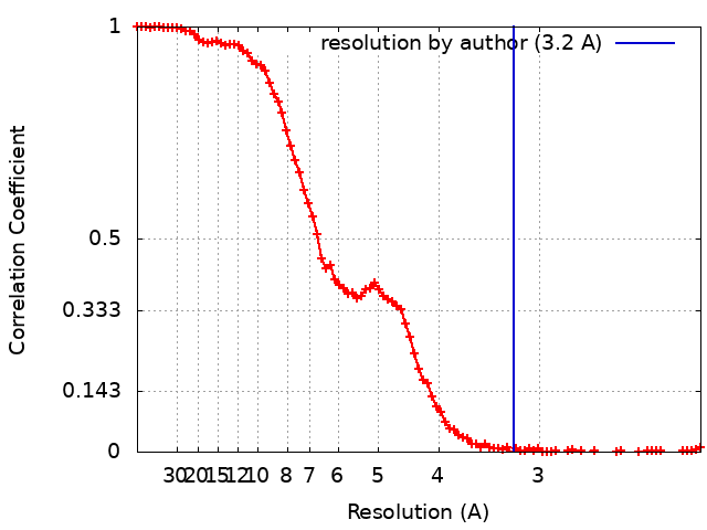 |

-Atomic model buiding 1
| Refinement | Space: REAL / Protocol: AB INITIO MODEL / Target criteria: correlation coefficient |
|---|---|
| Output model | PDB-7z11: |
