 Movie
Movie Controller

[English] 日本語
 Yorodumi
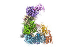
- PDB-7v6c: Structure of the Dicer-2-R2D2 heterodimer bound to small RNA duplex -
+ Open data

- Basic information
| Entry | Database: PDB / ID: 7v6c | ||||||
|---|---|---|---|---|---|---|---|
| Title | Structure of the Dicer-2-R2D2 heterodimer bound to small RNA duplex | ||||||
 Components Components |
| ||||||
 Keywords Keywords |  RNA BINDING PROTEIN / Ribonuclease / Double-stranded RNA-binding protein RNA BINDING PROTEIN / Ribonuclease / Double-stranded RNA-binding protein | ||||||
| Function / homology |  Function and homology information Function and homology informationfollicle cell of egg chamber stalk formation / regulatory ncRNA processing / bidentate ribonuclease III activity / positive regulation of Toll signaling pathway / MicroRNA (miRNA) biogenesis / Small interfering RNA (siRNA) biogenesis / PKR-mediated signaling / dsRNA transport / regulation of regulatory ncRNA processing / dosage compensation by hyperactivation of X chromosome ...follicle cell of egg chamber stalk formation / regulatory ncRNA processing / bidentate ribonuclease III activity / positive regulation of Toll signaling pathway / MicroRNA (miRNA) biogenesis / Small interfering RNA (siRNA) biogenesis / PKR-mediated signaling / dsRNA transport / regulation of regulatory ncRNA processing / dosage compensation by hyperactivation of X chromosome / global gene silencing by mRNA cleavage / ribonuclease III / deoxyribonuclease I activity / apoptotic DNA fragmentation / detection of virus / RISC-loading complex / regulatory ncRNA-mediated post-transcriptional gene silencing / RISC complex assembly / ribonuclease III activity / siRNA processing / siRNA binding / ATP-dependent activity, acting on RNA / positive regulation of innate immune response / RISC complex / heterochromatin formation / positive regulation of defense response to virus by host / locomotory behavior / mRNA 3'-UTR binding / helicase activity / cytoplasmic ribonucleoprotein granule / cellular response to virus / double-stranded RNA binding / defense response to virus / perinuclear region of cytoplasm / ATP hydrolysis activity / RNA binding / ATP binding / nucleus / cytoplasmSimilarity search - Function | ||||||
| Biological species |  Drosophila melanogaster (fruit fly) Drosophila melanogaster (fruit fly) | ||||||
| Method | ELECTRON MICROSCOPY / single particle reconstruction / cryo EM / Resolution: 3.3 Å | ||||||
 Authors Authors | Yamaguchi, S. / Nishizawa, T. / Kusakizako, T. / Yamashita, K. / Tomita, A. / Hirano, H. / Nishimasu, H. / Nureki, O. | ||||||
| Funding support |  Japan, 1items Japan, 1items
| ||||||
 Citation Citation | Journal: Nature / Year: 2022 Title: Structure of the Dicer-2-R2D2 heterodimer bound to a small RNA duplex. Authors: Sonomi Yamaguchi / Masahiro Naganuma / Tomohiro Nishizawa / Tsukasa Kusakizako / Yukihide Tomari / Hiroshi Nishimasu / Osamu Nureki / Abstract: In flies, Argonaute2 (Ago2) and small interfering RNA (siRNA) form an RNA-induced silencing complex to repress viral transcripts. The RNase III enzyme Dicer-2 associates with its partner protein R2D2 ...In flies, Argonaute2 (Ago2) and small interfering RNA (siRNA) form an RNA-induced silencing complex to repress viral transcripts. The RNase III enzyme Dicer-2 associates with its partner protein R2D2 and cleaves long double-stranded RNAs to produce 21-nucleotide siRNA duplexes, which are then loaded into Ago2 in a defined orientation. Here we report cryo-electron microscopy structures of the Dicer-2-R2D2 and Dicer-2-R2D2-siRNA complexes. R2D2 interacts with the helicase domain and the central linker of Dicer-2 to inhibit the promiscuous processing of microRNA precursors by Dicer-2. Notably, our structure represents the strand-selection state in the siRNA-loading process, and reveals that R2D2 asymmetrically recognizes the end of the siRNA duplex with the higher base-pairing stability, and the other end is exposed to the solvent and is accessible by Ago2. Our findings explain how R2D2 senses the thermodynamic asymmetry of the siRNA and facilitates the siRNA loading into Ago2 in a defined orientation, thereby determining which strand of the siRNA duplex is used by Ago2 as the guide strand for target silencing. | ||||||
| History |
|
- Structure visualization
| Structure viewer | Molecule: MolmilJmol/JSmol |
|---|
- Downloads & links
-Download
| PDBx/mmCIF format | 7v6c.cif.gz | 410.9 KB | Display | PDBx/mmCIF format |
|---|---|---|---|---|
| PDB format | pdb7v6c.ent.gz | 321.7 KB | Display | PDB format |
| PDBx/mmJSON format | 7v6c.json.gz | Tree view | PDBx/mmJSON format | |
| Others |  Other downloads Other downloads |
-Validation report
| Arichive directory | https://data.pdbj.org/pub/pdb/validation_reports/v6/7v6cftp://data.pdbj.org/pub/pdb/validation_reports/v6/7v6c | HTTPS FTP |
|---|
-Related structure data
| Related structure data | 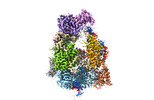 31742MC  7v6bC M: map data used to model this data C: citing same article ( |
|---|---|
| Similar structure data | |
| EM raw data | EMPIAR-11099 (Title: Structure of the Dicer-2-R2D2 heterodimer bound to a small RNA duplex Data size: 1.4 TB Data #1: Structure of the Dicer-2-R2D2 heterodimer [micrographs - multiframe] Data #2: Structure of the Dicer-2-R2D2 heterodimer bound to small RNA duplex [micrographs - multiframe]) |
-Links
 PDBj
PDBj


- Assembly
| Deposited unit | 
|
|---|---|
| 1 |
|
-Components
-Protein , 2 types, 2 molecules AB
| #1: Protein | / FI15132p1 Mass: 199309.000 Da / Num. of mol.: 1 / Mutation: M208L Source method: isolated from a genetically manipulated source Source: (gene. exp.) Drosophila melanogaster (fruit fly)Gene: Dcr-2, cg6493, Dcr, dcr, DCR-2, dcr-2, Dcr-2-RA, DCR2, Dcr2, dcr2, dDcr2, dic2, DICER, Dicer, dicer, DICER-2, dicer-2, Dicer2, dicer2, dmDcr-2, Dmel\CG6493, CG6493, Dmel_CG6493 Production host:  Spodoptera frugiperda (fall armyworm) Spodoptera frugiperda (fall armyworm)References: UniProt: A1ZAW0, deoxyribonuclease I, Hydrolases; Acting on ester bonds; Endoribonucleases producing 5'-phosphomonoesters, ribonuclease III, EC: 3.6.1.3 |
|---|---|
| #2: Protein | Mass: 39920.363 Da / Num. of mol.: 1 Source method: isolated from a genetically manipulated source Source: (gene. exp.) Drosophila melanogaster (fruit fly)Gene: r2d2, cg7138, Dmel\CG7138, R2D2, R2d2, CG7138, Dmel_CG7138 Production host: Spodoptera frugiperda (fall armyworm) / References: UniProt: Q9VLW8 |
-RNA chain , 4 types, 4 molecules EFCD
| #3: RNA chain | Mass: 6812.045 Da / Num. of mol.: 1 / Source method: obtained synthetically / Source: (synth.) Drosophila melanogaster (fruit fly) |
|---|---|
| #4: RNA chain | Mass: 6514.892 Da / Num. of mol.: 1 / Source method: obtained synthetically / Source: (synth.) Drosophila melanogaster (fruit fly) |
| #5: RNA chain | Mass: 7141.251 Da / Num. of mol.: 1 / Source method: obtained synthetically / Source: (synth.) Drosophila melanogaster (fruit fly) |
| #6: RNA chain | Mass: 6820.074 Da / Num. of mol.: 1 / Source method: obtained synthetically / Source: (synth.) Drosophila melanogaster (fruit fly) |
-Experimental details
-Experiment
| Experiment | Method: ELECTRON MICROSCOPY |
|---|---|
| EM experiment | Aggregation state: PARTICLE / 3D reconstruction method: single particle reconstruction |
- Sample preparation
| Component | Name: Dicer-2-R2D2-siRNA / Type: COMPLEX / Entity ID: all / Source: RECOMBINANT |
|---|---|
| Molecular weight | Experimental value: NO |
| Source (natural) | Organism: Drosophila melanogaster (fruit fly) |
| Source (recombinant) | Organism: Spodoptera frugiperda (fall armyworm) |
| Buffer solution | pH: 7.4 |
| Specimen | Embedding applied: NO / Shadowing applied: NO / Staining applied: NO / Vitrification applied: YES |
| Vitrification | Cryogen name: NITROGEN |
- Electron microscopy imaging
| Experimental equipment |  Model: Titan Krios / Image courtesy: FEI Company |
|---|---|
| Microscopy | Model: FEI TITAN KRIOS |
| Electron gun | Electron source: FIELD EMISSION GUN / Accelerating voltage: 300 kV / Illumination mode: FLOOD BEAM |
| Electron lens | Mode: BRIGHT FIELDBright-field microscopy |
| Image recording | Electron dose: 48 e/Å2 / Film or detector model: GATAN K3 (6k x 4k) |
- Processing
| Software | Name: REFMAC / Version: 5.8.0267 / Classification: refinement | ||||||||||||||||||||||||||||||||||||||||||||||||||||||||||||||||||||||||||||||||||||||||||||||||||||||||||
|---|---|---|---|---|---|---|---|---|---|---|---|---|---|---|---|---|---|---|---|---|---|---|---|---|---|---|---|---|---|---|---|---|---|---|---|---|---|---|---|---|---|---|---|---|---|---|---|---|---|---|---|---|---|---|---|---|---|---|---|---|---|---|---|---|---|---|---|---|---|---|---|---|---|---|---|---|---|---|---|---|---|---|---|---|---|---|---|---|---|---|---|---|---|---|---|---|---|---|---|---|---|---|---|---|---|---|---|
| CTF correction | Type: PHASE FLIPPING AND AMPLITUDE CORRECTION | ||||||||||||||||||||||||||||||||||||||||||||||||||||||||||||||||||||||||||||||||||||||||||||||||||||||||||
| 3D reconstruction | Resolution: 3.3 Å / Resolution method: FSC 0.143 CUT-OFF / Num. of particles: 144979 / Symmetry type: POINT | ||||||||||||||||||||||||||||||||||||||||||||||||||||||||||||||||||||||||||||||||||||||||||||||||||||||||||
| Refinement | Resolution: 3.3→165.75 Å / Cor.coef. Fo:Fc: 0.943 / SU B: 16.404 / SU ML: 0.268 / ESU R: 0.423 Stereochemistry target values: MAXIMUM LIKELIHOOD WITH PHASES Details: HYDROGENS HAVE BEEN ADDED IN THE RIDING POSITIONS
| ||||||||||||||||||||||||||||||||||||||||||||||||||||||||||||||||||||||||||||||||||||||||||||||||||||||||||
| Solvent computation | Solvent model: PARAMETERS FOR MASK CACLULATION | ||||||||||||||||||||||||||||||||||||||||||||||||||||||||||||||||||||||||||||||||||||||||||||||||||||||||||
| Displacement parameters | Biso mean: 101.99 Å2 | ||||||||||||||||||||||||||||||||||||||||||||||||||||||||||||||||||||||||||||||||||||||||||||||||||||||||||
| Refinement step | Cycle: 1 / Total: 15643 | ||||||||||||||||||||||||||||||||||||||||||||||||||||||||||||||||||||||||||||||||||||||||||||||||||||||||||
| Refine LS restraints |
|