 Movie
Movie Controller

[English] 日本語
 Yorodumi
- PDB-7tnv: Crystal Structure Of Human NADH-Cytochrome B5 Reductase T117S Mutant -
+ Open data

- Basic information
| Entry | Database: PDB / ID: 7tnv | ||||||
|---|---|---|---|---|---|---|---|
| Title | Crystal Structure Of Human NADH-Cytochrome B5 Reductase T117S Mutant | ||||||
 Components Components | NADH-cytochrome b5 reductase 3 soluble form | ||||||
 Keywords Keywords |  OXIDOREDUCTASE / FAD / B5R / Reductase / T117S OXIDOREDUCTASE / FAD / B5R / Reductase / T117S | ||||||
| Function / homology |  Function and homology informationnitric-oxide synthase complex / cytochrome-b5 reductase / Vitamin C (ascorbate) metabolism / cytochrome-b5 reductase activity, acting on NAD(P)H / Phase I - Functionalization of compounds / blood circulation / AMP binding / cholesterol biosynthetic process / hemoglobin complex / nitric oxide biosynthetic process ...nitric-oxide synthase complex / cytochrome-b5 reductase / Vitamin C (ascorbate) metabolism / cytochrome-b5 reductase activity, acting on NAD(P)H / Phase I - Functionalization of compounds / blood circulation / AMP binding / cholesterol biosynthetic process / hemoglobin complex / nitric oxide biosynthetic process / FAD binding / lipid droplet / mitochondrial membrane / ADP binding / NAD binding / azurophil granule lumen / mitochondrial outer membrane / Neutrophil degranulation / endoplasmic reticulum membrane / endoplasmic reticulum / mitochondrion / extracellular region / membrane / cytosol / cytoplasm Function and homology informationnitric-oxide synthase complex / cytochrome-b5 reductase / Vitamin C (ascorbate) metabolism / cytochrome-b5 reductase activity, acting on NAD(P)H / Phase I - Functionalization of compounds / blood circulation / AMP binding / cholesterol biosynthetic process / hemoglobin complex / nitric oxide biosynthetic process ...nitric-oxide synthase complex / cytochrome-b5 reductase / Vitamin C (ascorbate) metabolism / cytochrome-b5 reductase activity, acting on NAD(P)H / Phase I - Functionalization of compounds / blood circulation / AMP binding / cholesterol biosynthetic process / hemoglobin complex / nitric oxide biosynthetic process / FAD binding / lipid droplet / mitochondrial membrane / ADP binding / NAD binding / azurophil granule lumen / mitochondrial outer membrane / Neutrophil degranulation / endoplasmic reticulum membrane / endoplasmic reticulum / mitochondrion / extracellular region / membrane / cytosol / cytoplasmSimilarity search - Function | ||||||
| Biological species |  Homo sapiens (human) Homo sapiens (human) | ||||||
| Method | X-RAY DIFFRACTION / MOLECULAR REPLACEMENT / Resolution: 1.93 Å | ||||||
 Authors Authors | Zheng, A. / Thibodeau, P.H. | ||||||
| Funding support |  United States, 1items United States, 1items
| ||||||
 Citation Citation | Journal: to be published Title: Structure Of wild-type Human NADH-Cytochrome B5 Reductase and mutants Authors: Zheng, A. / Thibodeau, P.H. | ||||||
| History |
|
- Structure visualization
| Structure viewer | Molecule: MolmilJmol/JSmol |
|---|
- Downloads & links
-Download
| PDBx/mmCIF format | 7tnv.cif.gz | 92.8 KB | Display | PDBx/mmCIF format |
|---|---|---|---|---|
| PDB format | pdb7tnv.ent.gz | 55 KB | Display | PDB format |
| PDBx/mmJSON format | 7tnv.json.gz | Tree view | PDBx/mmJSON format | |
| Others |  Other downloads Other downloads |
-Validation report
| Arichive directory | https://data.pdbj.org/pub/pdb/validation_reports/tn/7tnvftp://data.pdbj.org/pub/pdb/validation_reports/tn/7tnv | HTTPS FTP |
|---|
-Related structure data
| Related structure data | 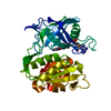 7thgC 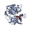 7tswC 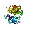 1umkS C: citing same article ( S: Starting model for refinement |
|---|---|
| Similar structure data |
-Links
 PDBj
PDBj


- Assembly
| Deposited unit | 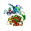
| ||||||||||||
|---|---|---|---|---|---|---|---|---|---|---|---|---|---|
| 1 |
| ||||||||||||
| Unit cell |
|
-Components
| #1: Protein | Mass: 31137.947 Da / Num. of mol.: 1 / Mutation: T117S Source method: isolated from a genetically manipulated source Source: (gene. exp.) Homo sapiens (human) / Gene: CYB5R3, DIA1 / Production host:  Escherichia coli (E. coli) / References: UniProt: P00387 Escherichia coli (E. coli) / References: UniProt: P00387 |
|---|---|
| #2: Chemical | ChemComp-FAD / Flavin adenine dinucleotide  Mass: 785.550 Da / Num. of mol.: 1 / Source method: obtained synthetically / Formula: C27H33N9O15P2 / Feature type: SUBJECT OF INVESTIGATION / Comment: FAD*YM Mass: 785.550 Da / Num. of mol.: 1 / Source method: obtained synthetically / Formula: C27H33N9O15P2 / Feature type: SUBJECT OF INVESTIGATION / Comment: FAD*YM |
| #3: Water | ChemComp-HOH / Water Mass: 18.015 Da / Num. of mol.: 271 / Source method: isolated from a natural source / Formula: H2O Mass: 18.015 Da / Num. of mol.: 271 / Source method: isolated from a natural source / Formula: H2O |
| Has ligand of interest | Y |
-Experimental details
-Experiment
| Experiment | Method: X-RAY DIFFRACTION / Number of used crystals: 1 |
|---|
- Sample preparation
| Crystal | Density Matthews: 2.48 Å3/Da / Density % sol: 50.35 % |
|---|---|
| Crystal grow | Temperature: 293 K / Method: vapor diffusion, sitting drop / Details: 0.1 M Tris-HCl pH 7.8, PEG 4K 10% |
-Data collection
| Diffraction | Mean temperature: 100 K / Serial crystal experiment: N |
|---|---|
| Diffraction source | Source: ROTATING ANODE / Type: RIGAKU FR-E SUPERBRIGHT / Wavelength: 1.54178 Å |
| Detector | Type: RIGAKU SATURN 944 / Detector: CCD / Date: Sep 27, 2017 |
| Radiation | Protocol: SINGLE WAVELENGTH / Monochromatic (M) / Laue (L): M / Scattering type: x-ray |
| Radiation wavelength | Wavelength: 1.54178 Å / Relative weight: 1 |
| Reflection | Resolution: 1.93→46.34 Å / Num. obs: 22991 / % possible obs: 99.8 % / Redundancy: 12.6 % / Rmerge(I) obs: 0.07 / Rrim(I) all: 0.073 / Net I/av σ(I): 23.58 / Net I/σ(I): 21.8 |
| Reflection shell | Resolution: 1.93→1.98 Å / Mean I/σ(I) obs: 2.55 / Num. unique obs: 1640 / CC1/2: 0.809 / Rrim(I) all: 1.602 |
- Processing
| Software |
| |||||||||||||||||||||||||||||||||||||||||||||||||||||||||||||||||||||||||||||||||||||||||||||||||||||||||
|---|---|---|---|---|---|---|---|---|---|---|---|---|---|---|---|---|---|---|---|---|---|---|---|---|---|---|---|---|---|---|---|---|---|---|---|---|---|---|---|---|---|---|---|---|---|---|---|---|---|---|---|---|---|---|---|---|---|---|---|---|---|---|---|---|---|---|---|---|---|---|---|---|---|---|---|---|---|---|---|---|---|---|---|---|---|---|---|---|---|---|---|---|---|---|---|---|---|---|---|---|---|---|---|---|---|---|
| Refinement | Method to determine structure: MOLECULAR REPLACEMENT Starting model: 1umk Resolution: 1.93→46.34 Å / SU ML: 0.2269 / Cross valid method: FREE R-VALUE / σ(F): 1.36 / Phase error: 22.5508 Stereochemistry target values: GeoStd + Monomer Library + CDL v1.2
| |||||||||||||||||||||||||||||||||||||||||||||||||||||||||||||||||||||||||||||||||||||||||||||||||||||||||
| Solvent computation | Shrinkage radii: 0.9 Å / VDW probe radii: 1.11 Å / Solvent model: FLAT BULK SOLVENT MODEL | |||||||||||||||||||||||||||||||||||||||||||||||||||||||||||||||||||||||||||||||||||||||||||||||||||||||||
| Displacement parameters | Biso mean: 35.03 Å2 | |||||||||||||||||||||||||||||||||||||||||||||||||||||||||||||||||||||||||||||||||||||||||||||||||||||||||
| Refinement step | Cycle: LAST / Resolution: 1.93→46.34 Å
| |||||||||||||||||||||||||||||||||||||||||||||||||||||||||||||||||||||||||||||||||||||||||||||||||||||||||
| Refine LS restraints |
| |||||||||||||||||||||||||||||||||||||||||||||||||||||||||||||||||||||||||||||||||||||||||||||||||||||||||
| LS refinement shell |
|
