 Movie
Movie Controller


+ Open data

- Basic information
| Entry | Database: PDB / ID: 1umk | ||||||
|---|---|---|---|---|---|---|---|
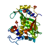
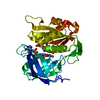
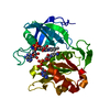
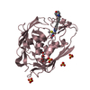
| Title | The Structure of Human Erythrocyte NADH-cytochrome b5 Reductase | ||||||
 Components Components | NADH-cytochrome b5 reductase | ||||||
 Keywords Keywords |  OXIDOREDUCTASE / flavoprotein / beta barrel / FAD-binding domain / NADH-binding domain OXIDOREDUCTASE / flavoprotein / beta barrel / FAD-binding domain / NADH-binding domain | ||||||
| Function / homology |  Function and homology informationnitric-oxide synthase complex / cytochrome-b5 reductase / Vitamin C (ascorbate) metabolism / cytochrome-b5 reductase activity, acting on NAD(P)H / Phase I - Functionalization of compounds / blood circulation / AMP binding / cholesterol biosynthetic process / hemoglobin complex / nitric oxide biosynthetic process ...nitric-oxide synthase complex / cytochrome-b5 reductase / Vitamin C (ascorbate) metabolism / cytochrome-b5 reductase activity, acting on NAD(P)H / Phase I - Functionalization of compounds / blood circulation / AMP binding / cholesterol biosynthetic process / hemoglobin complex / nitric oxide biosynthetic process / FAD binding / lipid droplet / mitochondrial membrane / ADP binding / NAD binding / azurophil granule lumen / mitochondrial outer membrane / Neutrophil degranulation / endoplasmic reticulum membrane / endoplasmic reticulum / mitochondrion / extracellular region / membrane / cytosol / cytoplasm Function and homology informationnitric-oxide synthase complex / cytochrome-b5 reductase / Vitamin C (ascorbate) metabolism / cytochrome-b5 reductase activity, acting on NAD(P)H / Phase I - Functionalization of compounds / blood circulation / AMP binding / cholesterol biosynthetic process / hemoglobin complex / nitric oxide biosynthetic process ...nitric-oxide synthase complex / cytochrome-b5 reductase / Vitamin C (ascorbate) metabolism / cytochrome-b5 reductase activity, acting on NAD(P)H / Phase I - Functionalization of compounds / blood circulation / AMP binding / cholesterol biosynthetic process / hemoglobin complex / nitric oxide biosynthetic process / FAD binding / lipid droplet / mitochondrial membrane / ADP binding / NAD binding / azurophil granule lumen / mitochondrial outer membrane / Neutrophil degranulation / endoplasmic reticulum membrane / endoplasmic reticulum / mitochondrion / extracellular region / membrane / cytosol / cytoplasmSimilarity search - Function | ||||||
| Biological species |  Homo sapiens (human) Homo sapiens (human) | ||||||
| Method | X-RAY DIFFRACTION / MIR / Resolution: 1.75 Å | ||||||
 Authors Authors | Bando, S. / Takano, T. / Yubisui, T. / Shirabe, K. / Takeshita, M. / Horii, C. / Nakagawa, A. | ||||||
 Citation Citation | Journal: Acta Crystallogr.,Sect.D / Year: 2004 Title: Structure of human erythrocyte NADH-cytochrome b5 reductase. Authors: Bando, S. / Takano, T. / Yubisui, T. / Shirabe, K. / Takeshita, M. / Nakagawa, A. | ||||||
| History |
|
- Structure visualization
| Structure viewer | Molecule: MolmilJmol/JSmol |
|---|
- Downloads & links
-Download
| PDBx/mmCIF format | 1umk.cif.gz | 88.3 KB | Display | PDBx/mmCIF format |
|---|---|---|---|---|
| PDB format | pdb1umk.ent.gz | 64.5 KB | Display | PDB format |
| PDBx/mmJSON format | 1umk.json.gz | Tree view | PDBx/mmJSON format | |
| Others |  Other downloads Other downloads |
-Validation report
| Arichive directory | https://data.pdbj.org/pub/pdb/validation_reports/um/1umkftp://data.pdbj.org/pub/pdb/validation_reports/um/1umk | HTTPS FTP |
|---|
-Related structure data
| Similar structure data |
|---|


-Links
 PDBj
PDBj


- Assembly
| Deposited unit | 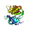
| ||||||||
|---|---|---|---|---|---|---|---|---|---|
| 1 |
| ||||||||
| Unit cell |
|
-Components
| #1: Protein | Mass: 31299.145 Da / Num. of mol.: 1 Source method: isolated from a genetically manipulated source Source: (gene. exp.) Homo sapiens (human) / Plasmid: pUC13 / Production host:  Escherichia coli (E. coli) / References: UniProt: P00387, cytochrome-b5 reductase Escherichia coli (E. coli) / References: UniProt: P00387, cytochrome-b5 reductase |
|---|---|
| #2: Chemical | ChemComp-FAD / Flavin adenine dinucleotide  Mass: 785.550 Da / Num. of mol.: 1 / Source method: obtained synthetically / Formula: C27H33N9O15P2 / Comment: FAD*YM Mass: 785.550 Da / Num. of mol.: 1 / Source method: obtained synthetically / Formula: C27H33N9O15P2 / Comment: FAD*YM |
| #3: Water | ChemComp-HOH / Water Mass: 18.015 Da / Num. of mol.: 753 / Source method: isolated from a natural source / Formula: H2O Mass: 18.015 Da / Num. of mol.: 753 / Source method: isolated from a natural source / Formula: H2O |
-Experimental details
-Experiment
| Experiment | Method: X-RAY DIFFRACTION / Number of used crystals: 1 |
|---|
- Sample preparation
| Crystal | Density Matthews: 1.98 Å3/Da / Density % sol: 37.45 % |
|---|---|
| Crystal grow | Temperature: 286 K / Method: vapor diffusion, sitting drop / pH: 7.6 Details: PEG 4000, potassium phosphate, pH 7.6, VAPOR DIFFUSION, SITTING DROP, temperature 286K |
-Data collection
| Diffraction | Mean temperature: 100 K |
|---|---|
| Diffraction source | Source: ROTATING ANODE / Type: RIGAKU ULTRAX 18 / Wavelength: 1.5418 Å |
| Detector | Type: RIGAKU RAXIS IV / Detector: IMAGE PLATE / Date: May 27, 2002 / Details: OSMIC Multilayer Optic mirror |
| Radiation | Monochromator: OSMIC mirror / Protocol: SINGLE WAVELENGTH / Monochromatic (M) / Laue (L): M / Scattering type: x-ray |
| Radiation wavelength | Wavelength: 1.5418 Å / Relative weight: 1 |
| Reflection | Resolution: 1.75→23.7 Å / Num. all: 32692 / Num. obs: 32639 / % possible obs: 99.4 % / Observed criterion σ(F): 3 / Observed criterion σ(I): -4 / Redundancy: 5.2 % / Biso Wilson estimate: 19.5 Å2 / Rmerge(I) obs: 0.056 / Rsym value: 0.05 / Net I/σ(I): 8.7 |
| Reflection shell | Resolution: 1.75→1.795 Å / Rmerge(I) obs: 0.143 / Mean I/σ(I) obs: 5.4 / % possible all: 98.3 |
- Processing
| Software |
| ||||||||||||||||||||||||||||||||||||||||||||||||||||||||||||||||||||||
|---|---|---|---|---|---|---|---|---|---|---|---|---|---|---|---|---|---|---|---|---|---|---|---|---|---|---|---|---|---|---|---|---|---|---|---|---|---|---|---|---|---|---|---|---|---|---|---|---|---|---|---|---|---|---|---|---|---|---|---|---|---|---|---|---|---|---|---|---|---|---|---|
| Refinement | Method to determine structure: MIR / Resolution: 1.75→23.7 Å / Cor.coef. Fo:Fc: 0.964 / Cor.coef. Fo:Fc free: 0.939 / SU B: 1.654 / SU ML: 0.055 / Isotropic thermal model: isotropic / Cross valid method: THROUGHOUT / σ(F): 3 / ESU R: 0.116 / ESU R Free: 0.115 Stereochemistry target values: MAXIMUM LIKELIHOOD WITH PHASES
| ||||||||||||||||||||||||||||||||||||||||||||||||||||||||||||||||||||||
| Solvent computation | Ion probe radii: 0.8 Å / Shrinkage radii: 0.8 Å / VDW probe radii: 1.4 Å / Solvent model: BABINET MODEL WITH MASK | ||||||||||||||||||||||||||||||||||||||||||||||||||||||||||||||||||||||
| Displacement parameters | Biso mean: 24.098 Å2
| ||||||||||||||||||||||||||||||||||||||||||||||||||||||||||||||||||||||
| Refinement step | Cycle: LAST / Resolution: 1.75→23.7 Å
| ||||||||||||||||||||||||||||||||||||||||||||||||||||||||||||||||||||||
| Refine LS restraints |
| ||||||||||||||||||||||||||||||||||||||||||||||||||||||||||||||||||||||
| LS refinement shell | Resolution: 1.75→1.795 Å / Total num. of bins used: 20
|
