 Movie
Movie Controller

[English] 日本語
 Yorodumi
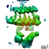
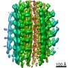
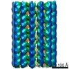
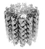
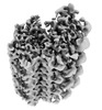
- PDB-7tns: Subpellicular microtubule from detergent-extract Toxoplasma gondi... -
+ Open data

- Basic information
| Entry | Database: PDB / ID: 7tns | ||||||||||||||||||
|---|---|---|---|---|---|---|---|---|---|---|---|---|---|---|---|---|---|---|---|
| Title | Subpellicular microtubule from detergent-extract Toxoplasma gondii cells | ||||||||||||||||||
 Components Components |
| ||||||||||||||||||
 Keywords Keywords | CELL INVASION /  parasites / Toxoplasma gondii / cytoskeleton / microtubules / tubulin parasites / Toxoplasma gondii / cytoskeleton / microtubules / tubulin | ||||||||||||||||||
| Function / homology |  Function and homology informationprotein-disulfide reductase / protein-disulfide reductase (NAD(P)H) activity / microtubule-based process / Hydrolases; Acting on acid anhydrides; Acting on GTP to facilitate cellular and subcellular movement / structural constituent of cytoskeleton / microtubule binding / microtubule / hydrolase activity / GTPase activity / GTP binding ...protein-disulfide reductase / protein-disulfide reductase (NAD(P)H) activity / microtubule-based process / Hydrolases; Acting on acid anhydrides; Acting on GTP to facilitate cellular and subcellular movement / structural constituent of cytoskeleton / microtubule binding / microtubule / hydrolase activity / GTPase activity / GTP binding / metal ion binding / cytoplasm Function and homology informationprotein-disulfide reductase / protein-disulfide reductase (NAD(P)H) activity / microtubule-based process / Hydrolases; Acting on acid anhydrides; Acting on GTP to facilitate cellular and subcellular movement / structural constituent of cytoskeleton / microtubule binding / microtubule / hydrolase activity / GTPase activity / GTP binding ...protein-disulfide reductase / protein-disulfide reductase (NAD(P)H) activity / microtubule-based process / Hydrolases; Acting on acid anhydrides; Acting on GTP to facilitate cellular and subcellular movement / structural constituent of cytoskeleton / microtubule binding / microtubule / hydrolase activity / GTPase activity / GTP binding / metal ion binding / cytoplasmSimilarity search - Function | ||||||||||||||||||
| Biological species |  Toxoplasma gondii (eukaryote) Toxoplasma gondii (eukaryote) | ||||||||||||||||||
| Method | ELECTRON MICROSCOPY / subtomogram averaging / cryo EM / Resolution: 6.7 Å | ||||||||||||||||||
 Authors Authors | Sun, S.Y. / Pintilie, G.D. / Chen, M. / Chiu, W. | ||||||||||||||||||
| Funding support |  United States, 5items United States, 5items
| ||||||||||||||||||
 Citation Citation | Journal: Proc Natl Acad Sci U S A / Year: 2022 Title: Cryo-ET of parasites gives subnanometer insight into tubulin-based structures. Authors: Stella Y Sun / Li-Av Segev-Zarko / Muyuan Chen / Grigore D Pintilie / Michael F Schmid / Steven J Ludtke / John C Boothroyd / Wah Chiu / Abstract: Tubulin is a conserved protein that polymerizes into different forms of filamentous structures in , an obligate intracellular parasite in the phylum Apicomplexa. Two key tubulin-containing ...Tubulin is a conserved protein that polymerizes into different forms of filamentous structures in , an obligate intracellular parasite in the phylum Apicomplexa. Two key tubulin-containing cytoskeletal components are subpellicular microtubules (SPMTs) and conoid fibrils (CFs). The SPMTs help maintain shape and gliding motility, while the CFs are implicated in invasion. Here, we use cryogenic electron tomography to determine the molecular structures of the SPMTs and CFs in vitrified intact and detergent-extracted parasites. Subvolume densities from detergent-extracted parasites yielded averaged density maps at subnanometer resolutions, and these were related back to their architecture in situ. An intralumenal spiral lines the interior of the 13-protofilament SPMTs, revealing a preferred orientation of these microtubules relative to the parasite's long axis. Each CF is composed of nine tubulin protofilaments that display a comma-shaped cross-section, plus additional associated components. Conoid protrusion, a crucial step in invasion, is associated with an altered pitch of each CF. The use of basic building blocks of protofilaments and different accessory proteins in one organism illustrates the versatility of tubulin to form two distinct types of assemblies, SPMTs and CFs. | ||||||||||||||||||
| History |
|
- Structure visualization
| Structure viewer | Molecule: MolmilJmol/JSmol |
|---|
- Downloads & links
-Download
| PDBx/mmCIF format | 7tns.cif.gz | 5.3 MB | Display | PDBx/mmCIF format |
|---|---|---|---|---|
| PDB format | pdb7tns.ent.gz | Display | PDB format | |
| PDBx/mmJSON format | 7tns.json.gz | Tree view | PDBx/mmJSON format | |
| Others |  Other downloads Other downloads |
-Validation report
| Arichive directory | https://data.pdbj.org/pub/pdb/validation_reports/tn/7tnsftp://data.pdbj.org/pub/pdb/validation_reports/tn/7tns | HTTPS FTP |
|---|
-Related structure data
| Related structure data | 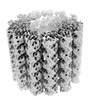 26019MC 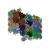 7tnqC  7tntC C: citing same article ( M: map data used to model this data |
|---|---|
| Similar structure data |
-Links
 PDBj
PDBj


- Assembly
| Deposited unit | 
|
|---|---|
| 1 |
|
-Components
| #1: Protein | Mass: 38817.699 Da / Num. of mol.: 24 / Source method: isolated from a natural source / Source: (natural) Toxoplasma gondii (eukaryote) / Strain: ATCC 50611 / Me49 / References: UniProt: S8F1Y1#2: Protein | Mass: 50166.645 Da / Num. of mol.: 26 / Source method: isolated from a natural source / Source: (natural) Toxoplasma gondii (eukaryote) / Strain: ME49 / References: UniProt: P10873#3: Protein | Mass: 50119.121 Da / Num. of mol.: 26 / Source method: isolated from a natural source / Source: (natural) Toxoplasma gondii (eukaryote) / Strain: ATCC 50611 / Me49 / References: UniProt: A0A125YWG5#4: Protein | Mass: 24939.332 Da / Num. of mol.: 24 / Source method: isolated from a natural source / Source: (natural) Toxoplasma gondii (eukaryote) / Strain: ATCC 50611 / Me49References: UniProt: A0A125YMM3, protein-disulfide reductase#5: Protein | | Mass: 21687.934 Da / Num. of mol.: 1 / Source method: isolated from a natural source / Source: (natural) Toxoplasma gondii (eukaryote) / Strain: ATCC 50611 / Me49 / References: UniProt: A0A125YFI4Has ligand of interest | N | |
|---|
-Experimental details
-Experiment
| Experiment | Method: ELECTRON MICROSCOPY |
|---|---|
| EM experiment | Aggregation state: FILAMENT / 3D reconstruction method: subtomogram averaging |
- Sample preparation
| Component | Name: microtubule complex of alpha-tubulin and beta-tubulin with intra-lumenal proteins Type: ORGANELLE OR CELLULAR COMPONENT / Entity ID: all / Source: NATURAL |
|---|---|
| Molecular weight | Experimental value: NO |
| Source (natural) | Organism: Toxoplasma gondii (eukaryote) / Strain: ME49 |
| Buffer solution | pH: 7.2 / Details: Phosphate-Buffered Saline |
| Specimen | Embedding applied: NO / Shadowing applied: NO / Staining applied: NO / Vitrification applied: YES |
| Specimen support | Grid material: COPPER / Grid mesh size: 200 divisions/in. / Grid type: Homemade |
| Vitrification | Instrument: LEICA EM GP / Cryogen name: ETHANE / Humidity: 90 % / Chamber temperature: 295 K |
- Electron microscopy imaging
| Experimental equipment |  Model: Titan Krios / Image courtesy: FEI Company |
|---|---|
| Microscopy | Model: FEI TITAN KRIOS |
| Electron gun | Electron source: FIELD EMISSION GUN / Accelerating voltage: 300 kV / Illumination mode: FLOOD BEAM |
| Electron lens | Mode: BRIGHT FIELDBright-field microscopy / Nominal magnification: 105000 X / Nominal defocus max: 5000 nm / Nominal defocus min: 1000 nm / Cs: 2.7 mm / C2 aperture diameter: 70 µm |
| Specimen holder | Cryogen: NITROGEN / Specimen holder model: FEI TITAN KRIOS AUTOGRID HOLDER |
| Image recording | Electron dose: 4.2 e/Å2 / Avg electron dose per subtomogram: 132 e/Å2 / Detector mode: COUNTING / Film or detector model: GATAN K2 SUMMIT (4k x 4k) |
| EM imaging optics | Energyfilter name: GIF Bioquantum / Energyfilter slit width: 20 eV |
| Image scans | Movie frames/image: 6 |
- Processing
| EM software |
| ||||||||||||||||
|---|---|---|---|---|---|---|---|---|---|---|---|---|---|---|---|---|---|
| CTF correction | Type: PHASE FLIPPING ONLY | ||||||||||||||||
| Helical symmerty | Angular rotation/subunit: 27.7 ° / Axial rise/subunit: 80 Å / Axial symmetry: C13 | ||||||||||||||||
| 3D reconstruction | Resolution: 6.7 Å / Resolution method: FSC 0.143 CUT-OFF / Num. of particles: 39122 / Algorithm: FOURIER SPACE / Symmetry type: HELICAL | ||||||||||||||||
| EM volume selection | Num. of tomograms: 90 / Num. of volumes extracted: 39122 | ||||||||||||||||
| Atomic model building | Protocol: RIGID BODY FIT | ||||||||||||||||
| Atomic model building | PDB-ID: 7MIZ |

