 Movie
Movie Controller

[English] 日本語
 Yorodumi
- EMDB-26018: The symmetry-released subpellicular microtubule map from detergen... -
+ Open data

- Basic information
| Entry | 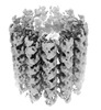 | ||||||||||||||||||
|---|---|---|---|---|---|---|---|---|---|---|---|---|---|---|---|---|---|---|---|
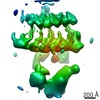
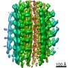
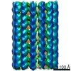
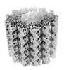
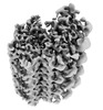
| Title | The symmetry-released subpellicular microtubule map from detergent-extracted Toxoplasma cells | ||||||||||||||||||
 Map data Map data | |||||||||||||||||||
 Sample Sample |
| ||||||||||||||||||
| Function / homology |  Function and homology information Function and homology information protein-disulfide reductase / protein-disulfide reductase (NAD(P)H) activity / microtubule-based process / Hydrolases; Acting on acid anhydrides; Acting on GTP to facilitate cellular and subcellular movement / structural constituent of cytoskeleton / microtubule binding / microtubule / hydrolase activity / GTPase activity / GTP binding ...protein-disulfide reductase / protein-disulfide reductase (NAD(P)H) activity / microtubule-based process / Hydrolases; Acting on acid anhydrides; Acting on GTP to facilitate cellular and subcellular movement / structural constituent of cytoskeleton / microtubule binding / microtubule / hydrolase activity / GTPase activity / GTP binding / metal ion binding / cytoplasm protein-disulfide reductase / protein-disulfide reductase (NAD(P)H) activity / microtubule-based process / Hydrolases; Acting on acid anhydrides; Acting on GTP to facilitate cellular and subcellular movement / structural constituent of cytoskeleton / microtubule binding / microtubule / hydrolase activity / GTPase activity / GTP binding ...protein-disulfide reductase / protein-disulfide reductase (NAD(P)H) activity / microtubule-based process / Hydrolases; Acting on acid anhydrides; Acting on GTP to facilitate cellular and subcellular movement / structural constituent of cytoskeleton / microtubule binding / microtubule / hydrolase activity / GTPase activity / GTP binding / metal ion binding / cytoplasmSimilarity search - Function | ||||||||||||||||||
| Biological species |  Toxoplasma gondii (eukaryote) Toxoplasma gondii (eukaryote) | ||||||||||||||||||
| Method | subtomogram averaging / cryo EM / Resolution: 8.4 Å | ||||||||||||||||||
 Authors Authors | Sun SY / Pintilie GD / Chen M | ||||||||||||||||||
| Funding support |  United States, 5 items United States, 5 items
| ||||||||||||||||||
 Citation Citation | Journal: Proc Natl Acad Sci U S A / Year: 2022 Title: Cryo-ET of parasites gives subnanometer insight into tubulin-based structures. Authors: Stella Y Sun / Li-Av Segev-Zarko / Muyuan Chen / Grigore D Pintilie / Michael F Schmid / Steven J Ludtke / John C Boothroyd / Wah Chiu / Abstract: Tubulin is a conserved protein that polymerizes into different forms of filamentous structures in , an obligate intracellular parasite in the phylum Apicomplexa. Two key tubulin-containing ...Tubulin is a conserved protein that polymerizes into different forms of filamentous structures in , an obligate intracellular parasite in the phylum Apicomplexa. Two key tubulin-containing cytoskeletal components are subpellicular microtubules (SPMTs) and conoid fibrils (CFs). The SPMTs help maintain shape and gliding motility, while the CFs are implicated in invasion. Here, we use cryogenic electron tomography to determine the molecular structures of the SPMTs and CFs in vitrified intact and detergent-extracted parasites. Subvolume densities from detergent-extracted parasites yielded averaged density maps at subnanometer resolutions, and these were related back to their architecture in situ. An intralumenal spiral lines the interior of the 13-protofilament SPMTs, revealing a preferred orientation of these microtubules relative to the parasite's long axis. Each CF is composed of nine tubulin protofilaments that display a comma-shaped cross-section, plus additional associated components. Conoid protrusion, a crucial step in invasion, is associated with an altered pitch of each CF. The use of basic building blocks of protofilaments and different accessory proteins in one organism illustrates the versatility of tubulin to form two distinct types of assemblies, SPMTs and CFs. | ||||||||||||||||||
| History |
|
- Structure visualization
| Supplemental images |
|---|

- Downloads & links
-EMDB archive
| Map data | emd_26018.map.gz | 10.8 MB | EMDB map data format | |
|---|---|---|---|---|
| Header (meta data) | emd-26018-v30.xmlemd-26018.xml | 17.8 KB 17.8 KB | Display Display | EMDB header |
| Images | 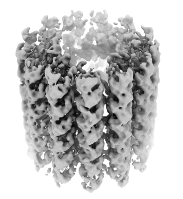 emd_26018.png emd_26018.png | 101.9 KB | ||
| Archive directory |  http://ftp.pdbj.org/pub/emdb/structures/EMD-26018ftp://ftp.pdbj.org/pub/emdb/structures/EMD-26018 http://ftp.pdbj.org/pub/emdb/structures/EMD-26018ftp://ftp.pdbj.org/pub/emdb/structures/EMD-26018 | HTTPS FTP |
-Related structure data
| Related structure data |  7tnqMC  7tnsC  7tntC M: atomic model generated by this map C: citing same article ( |
|---|---|
| Similar structure data |
-Links
| EMDB pages | EMDB (EBI/PDBe) / EMDataResource |
|---|---|
| Related items in Molecule of the Month |


-Map
| File | Download / File: emd_26018.map.gz / Format: CCP4 / Size: 38.4 MB / Type: IMAGE STORED AS FLOATING POINT NUMBER (4 BYTES) | ||||||||||||||||||||
|---|---|---|---|---|---|---|---|---|---|---|---|---|---|---|---|---|---|---|---|---|---|
| Voxel size | X=Y=Z: 2.0715 Å | ||||||||||||||||||||
| Density |
| ||||||||||||||||||||
| Symmetry | Space group: 1 | ||||||||||||||||||||
| Details | EMDB XML:
|
-Supplemental data
- Sample components
-Entire : microtubule complex of tubulin and intra-luminal proteins
| Entire | Name: microtubule complex of tubulin and intra-luminal proteins |
|---|---|
| Components |
|
-Supramolecule #1: microtubule complex of tubulin and intra-luminal proteins
| Supramolecule | Name: microtubule complex of tubulin and intra-luminal proteins type: organelle_or_cellular_component / ID: 1 / Parent: 0 / Macromolecule list: all |
|---|---|
| Source (natural) | Organism: Toxoplasma gondii (eukaryote) / Strain: ME49 |
-Macromolecule #1: Microtubule associated protein SPM1
| Macromolecule | Name: Microtubule associated protein SPM1 / type: protein_or_peptide / ID: 1 / Number of copies: 24 / Enantiomer: LEVO |
|---|---|
| Source (natural) | Organism: Toxoplasma gondii (eukaryote) / Strain: ATCC 50611 / Me49 |
| Molecular weight | Theoretical: 38.817699 KDa |
| Sequence | String: MSGGNSNTPK KLPSEEGSDY GYPQKPQKYL PKSEQAEPDY SACCKGNDAY KGASHGTVQF SHPEEAQKYA GAAAGAETIQ RGRERVAAD RQPRAAGDVP ARRLHLSDVD EAHRGQSPSR HPGYCVEELC TCGMHKCIPS RAPVPFTGST QYRQEFVPKP L PPPTQVSQ ...String: MSGGNSNTPK KLPSEEGSDY GYPQKPQKYL PKSEQAEPDY SACCKGNDAY KGASHGTVQF SHPEEAQKYA GAAAGAETIQ RGRERVAAD RQPRAAGDVP ARRLHLSDVD EAHRGQSPSR HPGYCVEELC TCGMHKCIPS RAPVPFTGST QYRQEFVPKP L PPPTQVSQ VTLPPSLPFE AESSYRTEFV AKPLPPPAKF SEVKLPPTLP FHGESAYRTD YVPKPLPEVA KPVEVKLPPT LP FNAQSCY RSEYVAKPLP PPVQTAEVKL PPSLPFEGST HYRDEFQVKP LPPATKVTEV KLPPSLPFDA TSMYRSDYVA KSN PICPVS KLPQYPAATY PQNHVFWDPD TKQWY |
-Macromolecule #2: Tubulin alpha chain
| Macromolecule | Name: Tubulin alpha chain / type: protein_or_peptide / ID: 2 / Number of copies: 26 / Enantiomer: LEVO |
|---|---|
| Source (natural) | Organism: Toxoplasma gondii (eukaryote) / Strain: ME49 |
| Molecular weight | Theoretical: 50.166645 KDa |
| Sequence | String: MREVISIHVG QAGIQIGNAC WELFCLEHGI QPDGQMPSDK TIGGGDDAFN TFFSETGAGK HVPRCVFLDL EPTVVDEVRT GTYRHLFHP EQLISGKEDA ANNFARGHYT IGKEIVDLSL DRIRKLADNC TGLQGFLMFN AVGGGTGSGL GCLLLERLSV D YGKKSKLN ...String: MREVISIHVG QAGIQIGNAC WELFCLEHGI QPDGQMPSDK TIGGGDDAFN TFFSETGAGK HVPRCVFLDL EPTVVDEVRT GTYRHLFHP EQLISGKEDA ANNFARGHYT IGKEIVDLSL DRIRKLADNC TGLQGFLMFN AVGGGTGSGL GCLLLERLSV D YGKKSKLN FCSWPSPQVS TAVVEPYNSV LSTHSLLEHT DVAVMLDNEA IYDICRRNLD IERPTYTNLN RLIAQVISSL TA SLRFDGA LNVDVTEFQT NLVPYPRIHF MLSSYAPIIS AEKAYHEQLS VAEITNSAFE PASMMAKCDP RHGKYMACCL MYR GDVVPK DVNAAVATIK TKRTIQFVDW CPTGFKCGIN YQPPTVVPGG DLAKVMRAVC MISNSTAIAE VFSRMDHKFD LMYA KRAFV HWYVGEGMEE GEFSEAREDL AALEKDYEEV GIETAEGEGE EEGYGDEY |
-Macromolecule #3: Tubulin beta chain
| Macromolecule | Name: Tubulin beta chain / type: protein_or_peptide / ID: 3 / Number of copies: 26 / Enantiomer: LEVO |
|---|---|
| Source (natural) | Organism: Toxoplasma gondii (eukaryote) / Strain: ATCC 50611 / Me49 |
| Molecular weight | Theoretical: 50.119121 KDa |
| Sequence | String: MREIVHVQGG QCGNQIGAKF WEVISDEHGI DPTGTYCGDS DLQLERINVF YNEATGGRFV PRAILMDLEP GTMDSVRAGP FGQLFRPDN FVFGQTGAGN NWAKGHYTEG AELIDSVLDV VRKEAEGCDC LQGFQITHSL GGGTGSGMGT LLISKVREEY P DRIMETFS ...String: MREIVHVQGG QCGNQIGAKF WEVISDEHGI DPTGTYCGDS DLQLERINVF YNEATGGRFV PRAILMDLEP GTMDSVRAGP FGQLFRPDN FVFGQTGAGN NWAKGHYTEG AELIDSVLDV VRKEAEGCDC LQGFQITHSL GGGTGSGMGT LLISKVREEY P DRIMETFS VFPSPKVSDT VVEPYNATLS VHQLVENADE VQVIDNEALY DICFRTLKLT TPTYGDLNHL VSAAMSGVTC CL RFPGQLN SDLRKLAVNL IPFPRLHFFL IGFAPLTSRG SQQYRALSVP ELTQQMFDAK NMMCASDPRH GRYLTASAMF RGR MSTKEV DEQMLNVQNK NSSYFVEWIP NNMKSSVCDI PPKGLKMSVT FVGNSTAIQE MFKRVSDQFT AMFRRKAFLH WYTG EGMDE MEFTEAESNM NDLVSEYQQY QDATAEEEGE FDEEEGEMGA EEGA |
-Macromolecule #4: PDI family protein
| Macromolecule | Name: PDI family protein / type: protein_or_peptide / ID: 4 / Number of copies: 20 / Enantiomer: LEVO / EC number: protein-disulfide reductase |
|---|---|
| Source (natural) | Organism: Toxoplasma gondii (eukaryote) / Strain: ATCC 50611 / Me49 |
| Molecular weight | Theoretical: 24.939332 KDa |
| Sequence | String: MSQPVFASPL NVEKRRLNEE RALMQAQKAG GEGVNIQLPP NYGDMDLILF PEGSLKNSNN TVIPQSHLKG KSVALYFADG ADPKCASLL PFLLNYYRTM NEGGANQKIE IIFVSLDRDR EAFESHRAHM PWLSIDLENP LTEILKRHFR VMKEYEVPTY G YGSRTGVP ...String: MSQPVFASPL NVEKRRLNEE RALMQAQKAG GEGVNIQLPP NYGDMDLILF PEGSLKNSNN TVIPQSHLKG KSVALYFADG ADPKCASLL PFLLNYYRTM NEGGANQKIE IIFVSLDRDR EAFESHRAHM PWLSIDLENP LTEILKRHFR VMKEYEVPTY G YGSRTGVP SVIVIGSDGR EAQFLPICSG LEEGDRALLR WDWRNTKFAS DQFHVRPTLL EQ |
-Macromolecule #5: PDI family protein
| Macromolecule | Name: PDI family protein / type: protein_or_peptide / ID: 5 / Number of copies: 4 / Enantiomer: LEVO |
|---|---|
| Source (natural) | Organism: Toxoplasma gondii (eukaryote) / Strain: ATCC 50611 / Me49 |
| Molecular weight | Theoretical: 21.687934 KDa |
| Sequence | String: MLAADCFFGP DVVKRTQQGN YVPVRPDHFA GVSVALFFAK AGHSKCAQIV PVVRQFYKTT NFSGEKAVIE IIYVSLDKDE QDFERVRAL MPWCSVEYKS CLRKKLIERY RVPNGELAFG TVRIPSTAIP LLIVIGPNGE EAGRMNFQQS DEFVLQRWDY R FNKWPGSA QRLRTLNDAT DPWKKRLPQN V |
-Experimental details
-Structure determination
| Method | cryo EM |
|---|---|
 Processing Processing | subtomogram averaging |
| Aggregation state | filament |
-Sample preparation
| Buffer | pH: 7.2 / Details: Phosphate Buffered Saline |
|---|---|
| Grid | Model: Homemade / Material: COPPER / Mesh: 200 / Support film - Material: CARBON / Support film - topology: LACEY / Pretreatment - Type: PLASMA CLEANING |
| Vitrification | Cryogen name: ETHANE / Chamber humidity: 90 % / Chamber temperature: 295 K / Instrument: LEICA EM GP |
- Electron microscopy
| Microscope | FEI TITAN KRIOS |
|---|---|
| Electron beam | Acceleration voltage: 300 kV / Electron source: FIELD EMISSION GUN |
| Electron optics | C2 aperture diameter: 70.0 µm / Illumination mode: FLOOD BEAM / Imaging mode: BRIGHT FIELDBright-field microscopy / Cs: 2.7 mm / Nominal defocus max: 5.0 µm / Nominal defocus min: 1.0 µm / Nominal magnification: 105000 |
| Specialist optics | Energy filter - Name: GIF Bioquantum / Energy filter - Slit width: 20 eV |
| Sample stage | Specimen holder model: FEI TITAN KRIOS AUTOGRID HOLDER / Cooling holder cryogen: NITROGEN |
| Image recording | Film or detector model: GATAN K2 SUMMIT (4k x 4k) / Detector mode: COUNTING / Average exposure time: 0.9 sec. / Average electron dose: 4.2 e/Å2 |
| Experimental equipment |  Model: Titan Krios / Image courtesy: FEI Company |
-Image processing
| Extraction | Number tomograms: 90 / Number images used: 39122 |
|---|---|
| Final angle assignment | Type: OTHER |
| Final reconstruction | Applied symmetry - Point group: C1 (asymmetric) / Algorithm: FOURIER SPACE / Resolution.type: BY AUTHOR / Resolution: 8.4 Å / Resolution method: FSC 0.143 CUT-OFF / Software - Name: EMAN2 / Number subtomograms used: 39122 |

