National Institutes of Health/National Institute of General Medical Sciences (NIH/NIGMS)
S10OD021600
United States
National Institutes of Health/National Institute of General Medical Sciences (NIH/NIGMS)
P41GM103832
United States
National Institutes of Health/National Institute of General Medical Sciences (NIH/NIGMS)
R01GM079429
United States
National Institutes of Health/National Institute of General Medical Sciences (NIH/NIGMS)
P01GM121203
United States
National Institutes of Health/National Institute of Mental Health (NIH/NIMH)
R21MH125285-01A1
United States
Citation
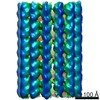
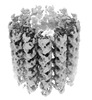
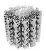
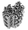
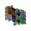
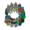
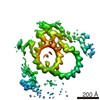
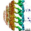
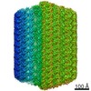
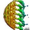
Journal: Proc Natl Acad Sci U S A / Year: 2022 Title: Cryo-ET of parasites gives subnanometer insight into tubulin-based structures. Authors: Stella Y Sun / Li-Av Segev-Zarko / Muyuan Chen / Grigore D Pintilie / Michael F Schmid / Steven J Ludtke / John C Boothroyd / Wah Chiu / Abstract: Tubulin is a conserved protein that polymerizes into different forms of filamentous structures in , an obligate intracellular parasite in the phylum Apicomplexa. Two key tubulin-containing ...Tubulin is a conserved protein that polymerizes into different forms of filamentous structures in , an obligate intracellular parasite in the phylum Apicomplexa. Two key tubulin-containing cytoskeletal components are subpellicular microtubules (SPMTs) and conoid fibrils (CFs). The SPMTs help maintain shape and gliding motility, while the CFs are implicated in invasion. Here, we use cryogenic electron tomography to determine the molecular structures of the SPMTs and CFs in vitrified intact and detergent-extracted parasites. Subvolume densities from detergent-extracted parasites yielded averaged density maps at subnanometer resolutions, and these were related back to their architecture in situ. An intralumenal spiral lines the interior of the 13-protofilament SPMTs, revealing a preferred orientation of these microtubules relative to the parasite's long axis. Each CF is composed of nine tubulin protofilaments that display a comma-shaped cross-section, plus additional associated components. Conoid protrusion, a crucial step in invasion, is associated with an altered pitch of each CF. The use of basic building blocks of protofilaments and different accessory proteins in one organism illustrates the versatility of tubulin to form two distinct types of assemblies, SPMTs and CFs.
History
Deposition
Jan 21, 2022
-
Header (metadata) release
Mar 2, 2022
-
Map release
Mar 2, 2022
-
Update
Jan 17, 2024
-
Current status
Jan 17, 2024
Processing site: RCSB / Status: Released
-
Structure visualization
Movie
Surface view with section colored by density value
In the structure databanks used in Yorodumi, some data are registered as the other names, "COVID-19 virus" and "2019-nCoV". Here are the details of the virus and the list of structure data.
Jan 31, 2019. EMDB accession codes are about to change! (news from PDBe EMDB page)
EMDB accession codes are about to change! (news from PDBe EMDB page)
The allocation of 4 digits for EMDB accession codes will soon come to an end. Whilst these codes will remain in use, new EMDB accession codes will include an additional digit and will expand incrementally as the available range of codes is exhausted. The current 4-digit format prefixed with “EMD-” (i.e. EMD-XXXX) will advance to a 5-digit format (i.e. EMD-XXXXX), and so on. It is currently estimated that the 4-digit codes will be depleted around Spring 2019, at which point the 5-digit format will come into force.
The EM Navigator/Yorodumi systems omit the EMD- prefix.
Related info.:Q: What is EMD? / ID/Accession-code notation in Yorodumi/EM Navigator
Yorodumi is a browser for structure data from EMDB, PDB, SASBDB, etc.
This page is also the successor to EM Navigator detail page, and also detail information page/front-end page for Omokage search.
The word "yorodu" (or yorozu) is an old Japanese word meaning "ten thousand". "mi" (miru) is to see.
Related info.:EMDB / PDB / SASBDB / Comparison of 3 databanks / Yorodumi Search / Aug 31, 2016. New EM Navigator & Yorodumi / Yorodumi Papers / Jmol/JSmol / Function and homology information / Changes in new EM Navigator and Yorodumi
 Movie
Movie Controller


 Open data
 Basic information
 Map data
 Sample
 Keywords
 parasites /
 Authors
 United States, 5 items
  Citation
 Structure visualization
 Movie viewer
 Downloads & links
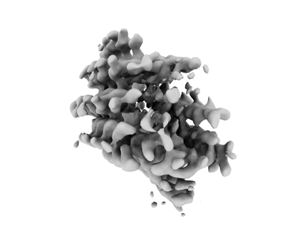 emd_26009.png
 http://ftp.pdbj.org/pub/emdb/structures/EMD-26009


 Sample components
 Processing
 Electron microscopy
