 Movie
Movie Controller

[English] 日本語
 Yorodumi
- PDB-7t5e: Neutron structure of Neurospora crassa Polysaccharide Monooxygena... -
+ Open data

- Basic information
| Entry | Database: PDB / ID: 7t5e | ||||||||||||
|---|---|---|---|---|---|---|---|---|---|---|---|---|---|
| Title | Neutron structure of Neurospora crassa Polysaccharide Monooxygenase 9D (NcLPMO9D) low pH vapor exchange | ||||||||||||
 Components Components | Lytic polysaccharide monooxygenase | ||||||||||||
 Keywords Keywords |  OXIDOREDUCTASE / LPMO / monooxygenase / PMO / metalloproteins OXIDOREDUCTASE / LPMO / monooxygenase / PMO / metalloproteins | ||||||||||||
| Function / homology | Auxiliary Activity family 9 / Auxiliary Activity family 9 (formerly GH61) / monooxygenase activity / hydrolase activity / extracellular region / metal ion binding / COPPER (II) ION / Lytic polysaccharide monooxygenase Function and homology information Function and homology information | ||||||||||||
| Biological species |  Neurospora crassa (fungus) Neurospora crassa (fungus) | ||||||||||||
| Method | X-RAY DIFFRACTION / NEUTRON DIFFRACTION / MOLECULAR REPLACEMENT / Resolution: 1.9 Å | ||||||||||||
 Authors Authors | Schroder, G.C. / Meilleur, F. | ||||||||||||
| Funding support |  United States, 3items United States, 3items
| ||||||||||||
 Citation Citation | Journal: Chem Sci / Year: 2022 Title: Capture of activated dioxygen intermediates at the copper-active site of a lytic polysaccharide monooxygenase. Authors: Schroder, G.C. / O'Dell, W.B. / Webb, S.P. / Agarwal, P.K. / Meilleur, F. #1: Journal: Acta Crystallogr F Struct Biol Commun / Year: 2021Title: Preliminary results of neutron and X-ray diffraction data collection on a lytic polysaccharide monooxygenase under reduced and acidic conditions. Authors: Schroder, G.C. / O'Dell, W.B. / Swartz, P.D. / Meilleur, F. | ||||||||||||
| History |
|
- Structure visualization
| Structure viewer | Molecule: MolmilJmol/JSmol |
|---|
- Downloads & links
-Download
| PDBx/mmCIF format | 7t5e.cif.gz | 210.7 KB | Display | PDBx/mmCIF format |
|---|---|---|---|---|
| PDB format | pdb7t5e.ent.gz | 172.1 KB | Display | PDB format |
| PDBx/mmJSON format | 7t5e.json.gz | Tree view | PDBx/mmJSON format | |
| Others |  Other downloads Other downloads |
-Validation report
| Arichive directory | https://data.pdbj.org/pub/pdb/validation_reports/t5/7t5eftp://data.pdbj.org/pub/pdb/validation_reports/t5/7t5e | HTTPS FTP |
|---|
-Related structure data
| Related structure data | 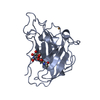 7t5cC  7t5dC 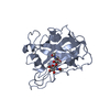 5tkhS C: citing same article ( S: Starting model for refinement |
|---|---|
| Similar structure data |
-Links
 PDBj
PDBj- Assembly
| Deposited unit | 
| ||||||||||
|---|---|---|---|---|---|---|---|---|---|---|---|
| 1 | 
| ||||||||||
| 2 | 
| ||||||||||
| Unit cell |
|
-Components
| #1: Protein | Mass: 23299.104 Da / Num. of mol.: 2 Source method: isolated from a genetically manipulated source Source: (gene. exp.) Neurospora crassa (fungus) / Gene: G15G9.090, GE21DRAFT_7469 / Production host: Komagataella pastoris (fungus) / Strain (production host): Superman5 / References: UniProt: Q8WZQ2#2: Polysaccharide | / Mass: 586.542 Da / Num. of mol.: 2 / Source method: obtained synthetically#3: Chemical | Copper  Mass: 63.546 Da / Num. of mol.: 2 / Source method: isolated from a natural source / Formula: Cu Mass: 63.546 Da / Num. of mol.: 2 / Source method: isolated from a natural source / Formula: Cu#4: Water | ChemComp-HOH / | Water Mass: 18.015 Da / Num. of mol.: 388 / Source method: isolated from a natural source / Formula: H2O Mass: 18.015 Da / Num. of mol.: 388 / Source method: isolated from a natural source / Formula: H2OHas ligand of interest | N | |
|---|
-Experimental details
-Experiment
| Experiment |
|
|---|
- Sample preparation
| Crystal | Density Matthews: 2.16 Å3/Da / Density % sol: 42.98 % / Description: Crystals form rectangular shapes. |
|---|---|
| Crystal grow | Temperature: 295 K / Method: vapor diffusion / pH: 4.4 / Details: PEG 3350, HEPES |
-Data collection
| Diffraction |
| |||||||||||||||||||||||||||
|---|---|---|---|---|---|---|---|---|---|---|---|---|---|---|---|---|---|---|---|---|---|---|---|---|---|---|---|---|
| Diffraction source |
| |||||||||||||||||||||||||||
| Detector |
| |||||||||||||||||||||||||||
| Radiation |
| |||||||||||||||||||||||||||
| Radiation wavelength |
| |||||||||||||||||||||||||||
| Reflection | Biso Wilson estimate: 17.17 Å2 / Entry-ID: 7T5E
| |||||||||||||||||||||||||||
| Reflection shell |
|
- Processing
| Software |
| |||||||||||||||||||||||||||||||||||||||||||||||||||||||||||||||||||||||||||||||||||||||||||||||||||||||||||||||||||||||||||||||||||||
|---|---|---|---|---|---|---|---|---|---|---|---|---|---|---|---|---|---|---|---|---|---|---|---|---|---|---|---|---|---|---|---|---|---|---|---|---|---|---|---|---|---|---|---|---|---|---|---|---|---|---|---|---|---|---|---|---|---|---|---|---|---|---|---|---|---|---|---|---|---|---|---|---|---|---|---|---|---|---|---|---|---|---|---|---|---|---|---|---|---|---|---|---|---|---|---|---|---|---|---|---|---|---|---|---|---|---|---|---|---|---|---|---|---|---|---|---|---|---|---|---|---|---|---|---|---|---|---|---|---|---|---|---|---|---|
| Refinement | SU ML: 0.18 / R Free selection details: Random selection / Cross valid method: FREE R-VALUE / Method to determine structure
| |||||||||||||||||||||||||||||||||||||||||||||||||||||||||||||||||||||||||||||||||||||||||||||||||||||||||||||||||||||||||||||||||||||
| Refinement step | Cycle: final / Resolution: 1.9→12.662 Å
| |||||||||||||||||||||||||||||||||||||||||||||||||||||||||||||||||||||||||||||||||||||||||||||||||||||||||||||||||||||||||||||||||||||
| Refine LS restraints |
| |||||||||||||||||||||||||||||||||||||||||||||||||||||||||||||||||||||||||||||||||||||||||||||||||||||||||||||||||||||||||||||||||||||
| LS refinement shell | Rfactor Rfree error: 0
|
