 Movie
Movie Controller

[English] 日本語
 Yorodumi
- PDB-7t5c: X-ray structure of Neurospora crassa Polysaccharide Monooxygenase... -
+ Open data

- Basic information
| Entry | Database: PDB / ID: 7t5c | ||||||||||||
|---|---|---|---|---|---|---|---|---|---|---|---|---|---|
| Title | X-ray structure of Neurospora crassa Polysaccharide Monooxygenase 9D (NcLPMO9D) at low pH | ||||||||||||
 Components Components | Lytic polysaccharide monooxygenase | ||||||||||||
 Keywords Keywords |  OXIDOREDUCTASE / LPMO / monooxygenase / PMO / metalloproteins / copper OXIDOREDUCTASE / LPMO / monooxygenase / PMO / metalloproteins / copper | ||||||||||||
| Function / homology | Auxiliary Activity family 9 / Auxiliary Activity family 9 (formerly GH61) / monooxygenase activity / hydrolase activity / extracellular region / metal ion binding / COPPER (II) ION / Lytic polysaccharide monooxygenase Function and homology information Function and homology information | ||||||||||||
| Biological species |  Neurospora crassa (fungus) Neurospora crassa (fungus) | ||||||||||||
| Method | X-RAY DIFFRACTION / MOLECULAR REPLACEMENT / Resolution: 1.5 Å | ||||||||||||
 Authors Authors | Schroder, G.C. / Meilleur, F. | ||||||||||||
| Funding support |  United States, 3items United States, 3items
| ||||||||||||
 Citation Citation | Journal: Chem Sci / Year: 2022 Title: Capture of activated dioxygen intermediates at the copper-active site of a lytic polysaccharide monooxygenase. Authors: Schroder, G.C. / O'Dell, W.B. / Webb, S.P. / Agarwal, P.K. / Meilleur, F. #1: Journal: Acta Crystallogr F Struct Biol Commun / Year: 2021Title: Preliminary results of neutron and X-ray diffraction data collection on a lytic polysaccharide monooxygenase under reduced and acidic conditions. Authors: Schroder, G.C. / O'Dell, W.B. / Swartz, P.D. / Meilleur, F. | ||||||||||||
| History |
|
- Structure visualization
| Structure viewer | Molecule: MolmilJmol/JSmol |
|---|
- Downloads & links
-Download
| PDBx/mmCIF format | 7t5c.cif.gz | 133.6 KB | Display | PDBx/mmCIF format |
|---|---|---|---|---|
| PDB format | pdb7t5c.ent.gz | 91.7 KB | Display | PDB format |
| PDBx/mmJSON format | 7t5c.json.gz | Tree view | PDBx/mmJSON format | |
| Others |  Other downloads Other downloads |
-Validation report
| Arichive directory | https://data.pdbj.org/pub/pdb/validation_reports/t5/7t5cftp://data.pdbj.org/pub/pdb/validation_reports/t5/7t5c | HTTPS FTP |
|---|
-Related structure data
| Related structure data |  7t5dC 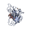 7t5eC 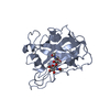 5tkhS C: citing same article ( S: Starting model for refinement |
|---|---|
| Similar structure data |
-Links
 PDBj
PDBj- Assembly
| Deposited unit | 
| ||||||||||||
|---|---|---|---|---|---|---|---|---|---|---|---|---|---|
| 1 | 
| ||||||||||||
| 2 | 
| ||||||||||||
| Unit cell |
|
-Components
| #1: Protein | Mass: 23299.104 Da / Num. of mol.: 2 Source method: isolated from a genetically manipulated source Source: (gene. exp.) Neurospora crassa (fungus) / Gene: G15G9.090, GE21DRAFT_7469 / Production host: Komagataella pastoris (fungus) / Strain (production host): SuperMan5 / References: UniProt: Q8WZQ2#2: Polysaccharide | 2-acetamido-2-deoxy-beta-D-glucopyranose-(1-4)-2-acetamido-2-deoxy-beta-D-glucopyranose | / Mass: 424.401 Da / Num. of mol.: 1Source method: isolated from a genetically manipulated source #3: Polysaccharide | alpha-D-mannopyranose-(1-4)-2-acetamido-2-deoxy-beta-D-glucopyranose-(1-4)-2-acetamido-2-deoxy-beta- ...alpha-D-mannopyranose-(1-4)-2-acetamido-2-deoxy-beta-D-glucopyranose-(1-4)-2-acetamido-2-deoxy-beta-D-glucopyranose | / Mass: 586.542 Da / Num. of mol.: 1Source method: isolated from a genetically manipulated source #4: Chemical | Copper  Mass: 63.546 Da / Num. of mol.: 2 / Source method: obtained synthetically / Formula: Cu / Feature type: SUBJECT OF INVESTIGATION Mass: 63.546 Da / Num. of mol.: 2 / Source method: obtained synthetically / Formula: Cu / Feature type: SUBJECT OF INVESTIGATION#5: Water | ChemComp-HOH / | Water Mass: 18.015 Da / Num. of mol.: 777 / Source method: isolated from a natural source / Formula: H2O Mass: 18.015 Da / Num. of mol.: 777 / Source method: isolated from a natural source / Formula: H2OHas ligand of interest | Y | |
|---|
-Experimental details
-Experiment
| Experiment | Method: X-RAY DIFFRACTION / Number of used crystals: 1 |
|---|
- Sample preparation
| Crystal | Density Matthews: 1.96 Å3/Da / Density % sol: 41.73 % / Description: Crystals form rectangular shapes. |
|---|---|
| Crystal grow | Temperature: 295 K / Method: vapor diffusion / pH: 4.4 / Details: PEG 3350, HEPES |
-Data collection
| Diffraction | Mean temperature: 100 K / Serial crystal experiment: N |
|---|---|
| Diffraction source | Source: ROTATING ANODE / Type: RIGAKU MICROMAX-007 / Wavelength: 1.54 Å |
| Detector | Type: DECTRIS EIGER R 4M / Detector: PIXEL / Date: Dec 4, 2018 |
| Radiation | Protocol: SINGLE WAVELENGTH / Monochromatic (M) / Laue (L): M / Scattering type: x-ray |
| Radiation wavelength | Wavelength: 1.54 Å / Relative weight: 1 |
| Reflection | Resolution: 1.5→12.61 Å / Num. obs: 62507 / % possible obs: 99.78 % / Redundancy: 8.2 % / Biso Wilson estimate: 11.79 Å2 / CC1/2: 0.992 / Rmerge(I) obs: 0.142 / Rrim(I) all: 0.1495 / Net I/σ(I): 62.56 |
| Reflection shell | Resolution: 1.5→1.55 Å / Redundancy: 4.5 % / Rmerge(I) obs: 0.2775 / Mean I/σ(I) obs: 7.14 / Num. unique obs: 6159 / CC1/2: 0.94 / Rrim(I) all: 0.3133 / % possible all: 99.97 |
- Processing
| Software |
| |||||||||||||||||||||||||||||||||||||||||||||||||||||||||||||||||||||||||||||||||||||||||||||||||||||||||||||||||||||||||||||||||||||||||||||||||||||||||||||||||
|---|---|---|---|---|---|---|---|---|---|---|---|---|---|---|---|---|---|---|---|---|---|---|---|---|---|---|---|---|---|---|---|---|---|---|---|---|---|---|---|---|---|---|---|---|---|---|---|---|---|---|---|---|---|---|---|---|---|---|---|---|---|---|---|---|---|---|---|---|---|---|---|---|---|---|---|---|---|---|---|---|---|---|---|---|---|---|---|---|---|---|---|---|---|---|---|---|---|---|---|---|---|---|---|---|---|---|---|---|---|---|---|---|---|---|---|---|---|---|---|---|---|---|---|---|---|---|---|---|---|---|---|---|---|---|---|---|---|---|---|---|---|---|---|---|---|---|---|---|---|---|---|---|---|---|---|---|---|---|---|---|---|---|
| Refinement | Method to determine structure: MOLECULAR REPLACEMENT Starting model: 5TKH Resolution: 1.5→12.61 Å / SU ML: 0.1158 / Cross valid method: FREE R-VALUE / σ(F): 1.16 / Phase error: 18.0267 / Stereochemistry target values: CDL v1.2
| |||||||||||||||||||||||||||||||||||||||||||||||||||||||||||||||||||||||||||||||||||||||||||||||||||||||||||||||||||||||||||||||||||||||||||||||||||||||||||||||||
| Solvent computation | Shrinkage radii: 0.9 Å / VDW probe radii: 1.11 Å / Solvent model: FLAT BULK SOLVENT MODEL | |||||||||||||||||||||||||||||||||||||||||||||||||||||||||||||||||||||||||||||||||||||||||||||||||||||||||||||||||||||||||||||||||||||||||||||||||||||||||||||||||
| Displacement parameters | Biso mean: 16.52 Å2 | |||||||||||||||||||||||||||||||||||||||||||||||||||||||||||||||||||||||||||||||||||||||||||||||||||||||||||||||||||||||||||||||||||||||||||||||||||||||||||||||||
| Refinement step | Cycle: LAST / Resolution: 1.5→12.61 Å
| |||||||||||||||||||||||||||||||||||||||||||||||||||||||||||||||||||||||||||||||||||||||||||||||||||||||||||||||||||||||||||||||||||||||||||||||||||||||||||||||||
| Refine LS restraints |
| |||||||||||||||||||||||||||||||||||||||||||||||||||||||||||||||||||||||||||||||||||||||||||||||||||||||||||||||||||||||||||||||||||||||||||||||||||||||||||||||||
| LS refinement shell |
|
