 Movie
Movie Controller

[English] 日本語
 Yorodumi
- PDB-7sej: Structure-based design of prefusion-stabilized human metapneumovi... -
+ Open data

- Basic information
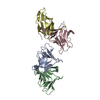
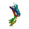
| Entry | Database: PDB / ID: 7sej | |||||||||
|---|---|---|---|---|---|---|---|---|---|---|
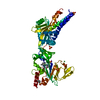
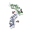
| Title | Structure-based design of prefusion-stabilized human metapneumovirus fusion proteins | |||||||||
 Components Components |
| |||||||||
 Keywords Keywords |  VIRAL PROTEIN / human metapneumovirus / fusion protein VIRAL PROTEIN / human metapneumovirus / fusion protein | |||||||||
| Function / homology | Precursor fusion glycoprotein F0, Paramyxoviridae / Fusion glycoprotein F0 / fusion of virus membrane with host plasma membrane / host cell plasma membrane / virion membrane / plasma membrane / Fusion glycoprotein F0 Function and homology information Function and homology information | |||||||||
| Biological species |  Human metapneumovirus Human metapneumovirus | |||||||||
| Method | X-RAY DIFFRACTION / SYNCHROTRON / MOLECULAR REPLACEMENT / Resolution: 2.51 Å | |||||||||
 Authors Authors | Hsieh, C.-L. / Rush, S.A. / McLellan, J.S. | |||||||||
| Funding support |  United States, 2items United States, 2items
| |||||||||
 Citation Citation | Journal: Nat Commun / Year: 2022 Title: Structure-based design of prefusion-stabilized human metapneumovirus fusion proteins. Authors: Hsieh, C.L. / Rush, S.A. / Palomo, C. / Chou, C.W. / Pickens, W. / Mas, V. / McLellan, J.S. | |||||||||
| History |
|
- Structure visualization
| Structure viewer | Molecule: MolmilJmol/JSmol |
|---|
- Downloads & links
-Download
| PDBx/mmCIF format | 7sej.cif.gz | 199 KB | Display | PDBx/mmCIF format |
|---|---|---|---|---|
| PDB format | pdb7sej.ent.gz | 141.3 KB | Display | PDB format |
| PDBx/mmJSON format | 7sej.json.gz | Tree view | PDBx/mmJSON format | |
| Others |  Other downloads Other downloads |
-Validation report
| Arichive directory | https://data.pdbj.org/pub/pdb/validation_reports/se/7sejftp://data.pdbj.org/pub/pdb/validation_reports/se/7sej | HTTPS FTP |
|---|
-Related structure data
| Related structure data |  7semC 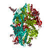 5wb0S S: Starting model for refinement C: citing same article ( |
|---|---|
| Similar structure data |


-Links
 PDBj
PDBj

- Assembly
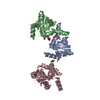
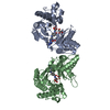
| Deposited unit | 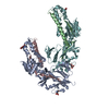
| ||||||||||||
|---|---|---|---|---|---|---|---|---|---|---|---|---|---|
| 1 | 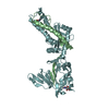
| ||||||||||||
| 2 | 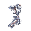
| ||||||||||||
| Unit cell |
|
-Components
| #1: Protein | Mass: 11015.444 Da / Num. of mol.: 2 Source method: isolated from a genetically manipulated source Source: (gene. exp.) Human metapneumovirus / Cell (production host): HEK293 / Production host:  Homo sapiens (human) / References: UniProt: H6X1Z0 Homo sapiens (human) / References: UniProt: H6X1Z0#2: Protein | Mass: 49456.418 Da / Num. of mol.: 2 Source method: isolated from a genetically manipulated source Details: the protein is cleaved into the two subunits (entities 1 and 2) as it transported through ER-Golgi Source: (gene. exp.) Human metapneumovirus / Cell (production host): HEK293 / Production host: Homo sapiens (human) / References: UniProt: H6X1Z0#3: Sugar | ChemComp-NAG / N-Acetylglucosamine  Type: D-saccharide, beta linking / Mass: 221.208 Da / Num. of mol.: 4 / Source method: obtained synthetically / Formula: C8H15NO6 Type: D-saccharide, beta linking / Mass: 221.208 Da / Num. of mol.: 4 / Source method: obtained synthetically / Formula: C8H15NO6#4: Water | ChemComp-HOH / | Water Mass: 18.015 Da / Num. of mol.: 136 / Source method: isolated from a natural source / Formula: H2O Mass: 18.015 Da / Num. of mol.: 136 / Source method: isolated from a natural source / Formula: H2OHas ligand of interest | N | |
|---|
-Experimental details
-Experiment
| Experiment | Method: X-RAY DIFFRACTION / Number of used crystals: 1 |
|---|
- Sample preparation
| Crystal | Density Matthews: 2.43 Å3/Da / Density % sol: 49.49 % |
|---|---|
| Crystal grow | Temperature: 298 K / Method: vapor diffusion, hanging drop / pH: 6 Details: 500 nl of hMPV F (10 mg/ml) with 500 nl of reservoir solution containing 0.1 M MES pH 6.0 and 12%(v/v) PEK 20k. |
-Data collection
| Diffraction | Mean temperature: 80 K / Serial crystal experiment: N |
|---|---|
| Diffraction source | Source: SYNCHROTRON / Site: APS / Beamline: 19-ID / Wavelength: 0.9792 Å |
| Detector | Type: DECTRIS PILATUS3 6M / Detector: PIXEL / Date: Nov 1, 2020 |
| Radiation | Protocol: SINGLE WAVELENGTH / Monochromatic (M) / Laue (L): M / Scattering type: x-ray |
| Radiation wavelength | Wavelength: 0.9792 Å / Relative weight: 1 |
| Reflection | Resolution: 2.5→45.4 Å / Num. obs: 35650 / % possible obs: 97.6 % / Redundancy: 3.2 % / Biso Wilson estimate: 40.37 Å2 / CC1/2: 0.994 / Rmerge(I) obs: 0.078 / Rpim(I) all: 0.073 / Net I/σ(I): 8.3 |
| Reflection shell | Resolution: 2.51→2.6 Å / Rmerge(I) obs: 0.038 / Mean I/σ(I) obs: 15.5 / Num. unique obs: 801 / CC1/2: 0.994 / Rpim(I) all: 0.037 |
- Processing
| Software |
| ||||||||||||||||||||||||||||||||||||||||||||||||||||||||||||||||||||||||||||||||||||||||||||||||||
|---|---|---|---|---|---|---|---|---|---|---|---|---|---|---|---|---|---|---|---|---|---|---|---|---|---|---|---|---|---|---|---|---|---|---|---|---|---|---|---|---|---|---|---|---|---|---|---|---|---|---|---|---|---|---|---|---|---|---|---|---|---|---|---|---|---|---|---|---|---|---|---|---|---|---|---|---|---|---|---|---|---|---|---|---|---|---|---|---|---|---|---|---|---|---|---|---|---|---|---|
| Refinement | Method to determine structure: MOLECULAR REPLACEMENT Starting model: 5wb0 Resolution: 2.51→45.31 Å / SU ML: 0.3247 / Cross valid method: FREE R-VALUE / σ(F): 1.34 / Phase error: 26.5753 Stereochemistry target values: GeoStd + Monomer Library + CDL v1.2
| ||||||||||||||||||||||||||||||||||||||||||||||||||||||||||||||||||||||||||||||||||||||||||||||||||
| Solvent computation | Shrinkage radii: 0.9 Å / VDW probe radii: 1.11 Å / Solvent model: FLAT BULK SOLVENT MODEL | ||||||||||||||||||||||||||||||||||||||||||||||||||||||||||||||||||||||||||||||||||||||||||||||||||
| Displacement parameters | Biso mean: 42.77 Å2 | ||||||||||||||||||||||||||||||||||||||||||||||||||||||||||||||||||||||||||||||||||||||||||||||||||
| Refinement step | Cycle: LAST / Resolution: 2.51→45.31 Å
| ||||||||||||||||||||||||||||||||||||||||||||||||||||||||||||||||||||||||||||||||||||||||||||||||||
| Refine LS restraints |
| ||||||||||||||||||||||||||||||||||||||||||||||||||||||||||||||||||||||||||||||||||||||||||||||||||
| LS refinement shell |
|
