 Movie
Movie Controller

[English] 日本語
 Yorodumi
- PDB-7s4r: Serial Macromolecular Crystallography at ALBA Synchrotron Light S... -
+ Open data

- Basic information
| Entry | Database: PDB / ID: 7s4r | |||||||||
|---|---|---|---|---|---|---|---|---|---|---|
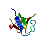
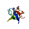
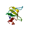
| Title | Serial Macromolecular Crystallography at ALBA Synchrotron Light Source - alpha Spectrin-SH3 domain | |||||||||
 Components Components | Spectrin alpha chain, non-erythrocytic 1 | |||||||||
 Keywords Keywords |  STRUCTURAL PROTEIN / Serial Crystallography STRUCTURAL PROTEIN / Serial Crystallography | |||||||||
| Function / homology |  Function and homology information Function and homology informationactin filament capping / costamere / cortical actin cytoskeleton / cell projection / actin filament binding / cell junction / actin cytoskeleton organization / calmodulin binding / calcium ion binding / plasma membraneSimilarity search - Function | |||||||||
| Biological species |  Gallus gallus (chicken) Gallus gallus (chicken) | |||||||||
| Method | X-RAY DIFFRACTION / SYNCHROTRON / MOLECULAR REPLACEMENT / Resolution: 2.1 Å | |||||||||
 Authors Authors | Martin-Garcia, J.M. / Botha, S. / Hu, H. / Jernigan, R. / Castellvi, A. / Lisova, S. / Gil, F. / Calisto, B. / Crespo, I. / Roy-Chowdbury, S. ...Martin-Garcia, J.M. / Botha, S. / Hu, H. / Jernigan, R. / Castellvi, A. / Lisova, S. / Gil, F. / Calisto, B. / Crespo, I. / Roy-Chowdbury, S. / Grieco, A. / Ketawala, G. / Weierstall, U. / Spence, J. / Fromme, P. / Zatsepin, N. / Boer, R. / Carpena, X. | |||||||||
| Funding support | 2items
| |||||||||
 Citation Citation | Journal: J.Synchrotron Radiat. / Year: 2022 Title: Serial macromolecular crystallography at ALBA Synchrotron Light Source. Authors: Martin-Garcia, J.M. / Botha, S. / Hu, H. / Jernigan, R. / Castellvi, A. / Lisova, S. / Gil, F. / Calisto, B. / Crespo, I. / Roy-Chowdhury, S. / Grieco, A. / Ketawala, G. / Weierstall, U. / ...Authors: Martin-Garcia, J.M. / Botha, S. / Hu, H. / Jernigan, R. / Castellvi, A. / Lisova, S. / Gil, F. / Calisto, B. / Crespo, I. / Roy-Chowdhury, S. / Grieco, A. / Ketawala, G. / Weierstall, U. / Spence, J. / Fromme, P. / Zatsepin, N. / Boer, D.R. / Carpena, X. | |||||||||
| History |
|
- Structure visualization
| Structure viewer | Molecule: MolmilJmol/JSmol |
|---|
- Downloads & links
-Download
| PDBx/mmCIF format | 7s4r.cif.gz | 26 KB | Display | PDBx/mmCIF format |
|---|---|---|---|---|
| PDB format | pdb7s4r.ent.gz | 14.3 KB | Display | PDB format |
| PDBx/mmJSON format | 7s4r.json.gz | Tree view | PDBx/mmJSON format | |
| Others |  Other downloads Other downloads |
-Validation report
| Arichive directory | https://data.pdbj.org/pub/pdb/validation_reports/s4/7s4rftp://data.pdbj.org/pub/pdb/validation_reports/s4/7s4r | HTTPS FTP |
|---|
-Related structure data
| Related structure data |  7s4wC  7s4yC  7s4zC  7s50C 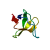 4f17S S: Starting model for refinement C: citing same article ( |
|---|---|
| Similar structure data |


-Links
 PDBj
PDBj- Assembly
| Deposited unit | 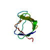
| ||||||||
|---|---|---|---|---|---|---|---|---|---|
| 1 |
| ||||||||
| Unit cell |
|
-Components
| #1: Protein | Mass: 6566.512 Da / Num. of mol.: 1 Source method: isolated from a genetically manipulated source Source: (gene. exp.) Gallus gallus (chicken) / Gene: SPTAN1, SPTA2 / Production host:  Escherichia coli BL21(DE3) (bacteria) / Strain (production host): BL21(DE3) / References: UniProt: P07751 Escherichia coli BL21(DE3) (bacteria) / Strain (production host): BL21(DE3) / References: UniProt: P07751 |
|---|---|
| #2: Water | ChemComp-HOH / Water Mass: 18.015 Da / Num. of mol.: 5 / Source method: isolated from a natural source / Formula: H2O Mass: 18.015 Da / Num. of mol.: 5 / Source method: isolated from a natural source / Formula: H2O |
-Experimental details
-Experiment
| Experiment | Method: X-RAY DIFFRACTION / Number of used crystals: 1 |
|---|
- Sample preparation
| Crystal | Density Matthews: 2.82 Å3/Da / Density % sol: 56.35 % |
|---|---|
| Crystal grow | Temperature: 277 K / Method: evaporation Details: Crystals were obtained by applying several concentration/dilution steps with milli-Q water inside standard Amicon Ultra 0.5 ml centrifugal filter (3 kDa cutoff). A crystalline slurry ...Details: Crystals were obtained by applying several concentration/dilution steps with milli-Q water inside standard Amicon Ultra 0.5 ml centrifugal filter (3 kDa cutoff). A crystalline slurry appeared when most citric acid buffer was removed |
-Data collection
| Diffraction | Mean temperature: 298 K / Serial crystal experiment: Y |
|---|---|
| Diffraction source | Source: SYNCHROTRON / Site: ALBA  / Beamline: XALOC / Wavelength: 0.97918 Å / Beamline: XALOC / Wavelength: 0.97918 Å |
| Detector | Type: DECTRIS PILATUS 6M / Detector: PIXEL / Date: Sep 6, 2018 |
| Radiation | Protocol: SINGLE WAVELENGTH / Monochromatic (M) / Laue (L): M / Scattering type: x-ray |
| Radiation wavelength | Wavelength: 0.97918 Å / Relative weight: 1 |
| Reflection | Resolution: 2.1→42.6 Å / Num. obs: 4682 / % possible obs: 100 % / Redundancy: 245 % / Biso Wilson estimate: 50.3 Å2 / CC1/2: 0.99 / CC star: 0.9975 / Net I/σ(I): 6.5 |
| Reflection shell | Resolution: 2.1→2.17 Å / Redundancy: 57 % / Mean I/σ(I) obs: 0.3 / Num. unique obs: 454 / CC1/2: 0.204 / CC star: 0.5817 / % possible all: 100 |
| Serial crystallography sample delivery | Description: LCP injection / Method: injection |
| Serial crystallography sample delivery injection | Injector diameter: 5.0E-5 µm |
| Serial crystallography data reduction | Frames failed index: 1145735 / Lattices indexed: 13039 |
- Processing
| Software |
| |||||||||||||||||||||||||||||||||||||||||||||||||||||||||||||||||||||||||||||||||||||||||||||||||||||||||
|---|---|---|---|---|---|---|---|---|---|---|---|---|---|---|---|---|---|---|---|---|---|---|---|---|---|---|---|---|---|---|---|---|---|---|---|---|---|---|---|---|---|---|---|---|---|---|---|---|---|---|---|---|---|---|---|---|---|---|---|---|---|---|---|---|---|---|---|---|---|---|---|---|---|---|---|---|---|---|---|---|---|---|---|---|---|---|---|---|---|---|---|---|---|---|---|---|---|---|---|---|---|---|---|---|---|---|
| Refinement | Method to determine structure: MOLECULAR REPLACEMENT Starting model: 4F17 Resolution: 2.1→32.64 Å / Cor.coef. Fo:Fc: 0.971 / Cor.coef. Fo:Fc free: 0.961 / SU B: 8.696 / SU ML: 0.191 / Cross valid method: THROUGHOUT / ESU R: 0.195 / ESU R Free: 0.177 / Stereochemistry target values: MAXIMUM LIKELIHOOD Details: HYDROGENS HAVE BEEN ADDED IN THE RIDING POSITIONS U VALUES : REFINED INDIVIDUALLY
| |||||||||||||||||||||||||||||||||||||||||||||||||||||||||||||||||||||||||||||||||||||||||||||||||||||||||
| Solvent computation | Ion probe radii: 0.9 Å / Shrinkage radii: 0.9 Å / VDW probe radii: 1.2 Å / Solvent model: MASK | |||||||||||||||||||||||||||||||||||||||||||||||||||||||||||||||||||||||||||||||||||||||||||||||||||||||||
| Displacement parameters | Biso mean: 62.257 Å2
| |||||||||||||||||||||||||||||||||||||||||||||||||||||||||||||||||||||||||||||||||||||||||||||||||||||||||
| Refinement step | Cycle: LAST / Resolution: 2.1→32.64 Å
| |||||||||||||||||||||||||||||||||||||||||||||||||||||||||||||||||||||||||||||||||||||||||||||||||||||||||
| Refine LS restraints |
| |||||||||||||||||||||||||||||||||||||||||||||||||||||||||||||||||||||||||||||||||||||||||||||||||||||||||
| LS refinement shell | Resolution: 2.101→2.155 Å / Total num. of bins used: 20
|
