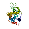
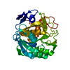
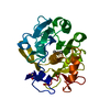
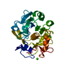
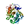
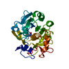
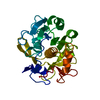
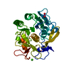
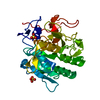
ProteinaseK / / Endopeptidase K / Tritirachium alkaline proteinase
Mass: 28958.791 Da / Num. of mol.: 1 Source method: isolated from a genetically manipulated source Source: (gene. exp.) Parengyodontium album (fungus) / Gene: PROK / Production host: Escherichia coli (E. coli) / References: UniProt: P06873, peptidase K
Resolution: 1.9→44.21 Å / Cor.coef. Fo:Fc: 0.974 / Cor.coef. Fo:Fc free: 0.96 / SU B: 6.602 / SU ML: 0.087 / Cross valid method: THROUGHOUT / ESU R: 0.138 / ESU R Free: 0.124 / Stereochemistry target values: MAXIMUM LIKELIHOOD Details: U VALUES : WITH TLS ADDED HYDROGENS HAVE BEEN ADDED IN THE RIDING POSITIONS U VALUES : RESIDUAL ONLY
Rfactor
Num. reflection
% reflection
Selection details
Rfree
0.19559
1071
5.1 %
RANDOM
Rwork
0.16508
-
-
-
obs
0.16664
19879
99.99 %
-
Solvent computation
Ion probe radii: 0.7 Å / Shrinkage radii: 0.7 Å / VDW probe radii: 1.2 Å / Solvent model: MASK
Displacement parameters
Biso mean: 30.94 Å2
Baniso -1
Baniso -2
Baniso -3
1-
0.09 Å2
-0 Å2
-0 Å2
2-
-
0.09 Å2
-0 Å2
3-
-
-
-0.18 Å2
Refinement step
Cycle: LAST / Resolution: 1.9→44.21 Å
Protein
Nucleic acid
Ligand
Solvent
Total
Num. atoms
2031
0
6
89
2126
Refine LS restraints
Refine-ID
Type
Dev ideal
Dev ideal target
Number
X-RAY DIFFRACTION
r_bond_refined_d
0.013
0.017
2075
X-RAY DIFFRACTION
r_bond_other_d
0.001
0.019
1854
X-RAY DIFFRACTION
r_angle_refined_deg
1.513
1.84
2820
X-RAY DIFFRACTION
r_angle_other_deg
1.196
2.664
4260
X-RAY DIFFRACTION
r_dihedral_angle_1_deg
6.283
5
278
X-RAY DIFFRACTION
r_dihedral_angle_2_deg
34.812
21.979
96
X-RAY DIFFRACTION
r_dihedral_angle_3_deg
12.819
15
299
X-RAY DIFFRACTION
r_dihedral_angle_4_deg
19.804
15
12
X-RAY DIFFRACTION
r_chiral_restr
0.088
0.2
312
X-RAY DIFFRACTION
r_gen_planes_refined
0.006
0.02
2461
X-RAY DIFFRACTION
r_gen_planes_other
0.001
0.02
495
X-RAY DIFFRACTION
r_mcbond_it
0.908
2.258
1115
X-RAY DIFFRACTION
r_mcbond_other
0.907
2.257
1114
X-RAY DIFFRACTION
r_mcangle_it
1.34
3.382
1392
X-RAY DIFFRACTION
r_mcangle_other
1.339
3.383
1393
X-RAY DIFFRACTION
r_scbond_it
1.637
2.513
960
X-RAY DIFFRACTION
r_scbond_other
1.635
2.504
958
X-RAY DIFFRACTION
r_scangle_other
2.672
3.67
1426
X-RAY DIFFRACTION
r_long_range_B_refined
3.957
27.394
2311
X-RAY DIFFRACTION
r_long_range_B_other
3.954
27.265
2300
LS refinement shell
Resolution: 1.9→1.949 Å / Total num. of bins used: 20
Rfactor
Num. reflection
% reflection
Rfree
0.516
79
-
Rwork
0.508
1431
-
obs
-
-
100 %
Refinement TLS params.
Method: refined / Origin x: 16.555 Å / Origin y: -12.766 Å / Origin z: -0.178 Å
11
12
13
21
22
23
31
32
33
T
0.0245 Å2
-0.0001 Å2
-0.0003 Å2
-
0.0007 Å2
-0.001 Å2
-
-
0.0349 Å2
L
1.2474 °2
-0.1246 °2
-0.0731 °2
-
1.3194 °2
0.0828 °2
-
-
0.8574 °2
S
-0.0062 Å °
-0.0007 Å °
0.0037 Å °
0.0852 Å °
0.0155 Å °
-0.0375 Å °
0.0338 Å °
-0.0187 Å °
-0.0094 Å °
+
About Yorodumi
-
News
-
Feb 9, 2022. New format data for meta-information of EMDB entries
New format data for meta-information of EMDB entries
Version 3 of the EMDB header file is now the official format.
The previous official version 1.9 will be removed from the archive.
In the structure databanks used in Yorodumi, some data are registered as the other names, "COVID-19 virus" and "2019-nCoV". Here are the details of the virus and the list of structure data.
Jan 31, 2019. EMDB accession codes are about to change! (news from PDBe EMDB page)
EMDB accession codes are about to change! (news from PDBe EMDB page)
The allocation of 4 digits for EMDB accession codes will soon come to an end. Whilst these codes will remain in use, new EMDB accession codes will include an additional digit and will expand incrementally as the available range of codes is exhausted. The current 4-digit format prefixed with “EMD-” (i.e. EMD-XXXX) will advance to a 5-digit format (i.e. EMD-XXXXX), and so on. It is currently estimated that the 4-digit codes will be depleted around Spring 2019, at which point the 5-digit format will come into force.
The EM Navigator/Yorodumi systems omit the EMD- prefix.
Related info.:Q: What is EMD? / ID/Accession-code notation in Yorodumi/EM Navigator
Yorodumi is a browser for structure data from EMDB, PDB, SASBDB, etc.
This page is also the successor to EM Navigator detail page, and also detail information page/front-end page for Omokage search.
The word "yorodu" (or yorozu) is an old Japanese word meaning "ten thousand". "mi" (miru) is to see.
Related info.:EMDB / PDB / SASBDB / Comparison of 3 databanks / Yorodumi Search / Aug 31, 2016. New EM Navigator & Yorodumi / Yorodumi Papers / Jmol/JSmol / Function and homology information / Changes in new EM Navigator and Yorodumi
 Movie
Movie Controller

 Yorodumi
 Open data
 Basic information
 Components
 Keywords
 Function and homology information
 Parengyodontium album (fungus)
 Authors
 Citation
 Structure visualization
 Downloads & links
 Other downloads
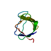


 PDBj
PDBj

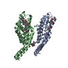
 Assembly


 Mass: 62.005 Da / Num. of mol.: 1 / Source method: obtained synthetically / Formula: NO3
 Mass: 40.078 Da / Num. of mol.: 2 / Source method: obtained synthetically / Formula: Ca
 Mass: 18.015 Da / Num. of mol.: 89 / Source method: isolated from a natural source / Formula: H2O
 Sample preparation
 / Beamline: XALOC / Wavelength: 0.97918 Å
 Processing