 Movie
Movie Controller

[English] 日本語
 Yorodumi
- PDB-7s2z: Crystal structure of the E100A mutant TIR domain from the grapevi... -
+ Open data

- Basic information
| Entry | Database: PDB / ID: 7s2z | |||||||||
|---|---|---|---|---|---|---|---|---|---|---|
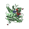
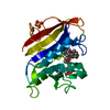
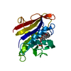
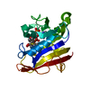
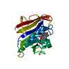
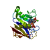
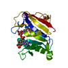
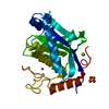
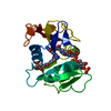
| Title | Crystal structure of the E100A mutant TIR domain from the grapevine disease resistance protein RUN1 bound to NAD | |||||||||
 Components Components | Disease resistance protein RUN1 | |||||||||
 Keywords Keywords | SIGNALING PROTEIN / HYDROLASE / NAD+ Hydrolase / Signalling Protein / TIR domain / NAD+ nucleosidase activity | |||||||||
| Function / homology |  Function and homology information Function and homology informationNAD catabolic process / NAD+ nucleosidase activity / ADP-ribosyl cyclase/cyclic ADP-ribose hydrolase / NAD+ nucleotidase, cyclic ADP-ribose generating / NADP+ nucleosidase activity / positive regulation of programmed cell death / Hydrolases; Glycosylases; Hydrolysing N-glycosyl compounds / defense response to fungus / ADP binding / signal transduction ...NAD catabolic process / NAD+ nucleosidase activity / ADP-ribosyl cyclase/cyclic ADP-ribose hydrolase / NAD+ nucleotidase, cyclic ADP-ribose generating / NADP+ nucleosidase activity / positive regulation of programmed cell death / Hydrolases; Glycosylases; Hydrolysing N-glycosyl compounds / defense response to fungus / ADP binding / signal transduction / nucleus / cytoplasmSimilarity search - Function | |||||||||
| Biological species |  Vitis rotundifolia (fox grape) Vitis rotundifolia (fox grape) | |||||||||
| Method | X-RAY DIFFRACTION / SYNCHROTRON / MOLECULAR REPLACEMENT / Resolution: 2.35 Å | |||||||||
 Authors Authors | Burdett, H. / Kobe, B. | |||||||||
| Funding support |  Australia, 2items Australia, 2items
| |||||||||
 Citation Citation | Journal: Biorxiv / Year: 2021 Title: Self-association configures the NAD + -binding site of plant NLR TIR domains Authors: Burdett, H. / Hu, X. / Rank, M.X. / Maruta, N. / Kobe, B. | |||||||||
| History |
|
- Structure visualization
| Structure viewer | Molecule: MolmilJmol/JSmol |
|---|
- Downloads & links
-Download
| PDBx/mmCIF format | 7s2z.cif.gz | 160.5 KB | Display | PDBx/mmCIF format |
|---|---|---|---|---|
| PDB format | pdb7s2z.ent.gz | 126.3 KB | Display | PDB format |
| PDBx/mmJSON format | 7s2z.json.gz | Tree view | PDBx/mmJSON format | |
| Others |  Other downloads Other downloads |
-Validation report
| Arichive directory | https://data.pdbj.org/pub/pdb/validation_reports/s2/7s2zftp://data.pdbj.org/pub/pdb/validation_reports/s2/7s2z | HTTPS FTP |
|---|
-Related structure data
| Related structure data | 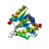 7rtsC 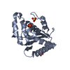 7rx1C 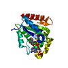 6o0wS S: Starting model for refinement C: citing same article ( |
|---|---|
| Similar structure data |

-Links
 PDBj
PDBj


- Assembly
| Deposited unit | 
| ||||||||||
|---|---|---|---|---|---|---|---|---|---|---|---|
| 1 | 
| ||||||||||
| 2 | 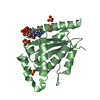
| ||||||||||
| 3 | 
| ||||||||||
| 4 | 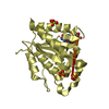
| ||||||||||
| Unit cell |
|
-Components
| #1: Protein | / NAD(+) hydrolase RUN1 / NADP(+) hydrolase RUN1 / Resistance to Uncinula necator protein / MrRUN1 Mass: 20655.346 Da / Num. of mol.: 4 / Mutation: E100A Source method: isolated from a genetically manipulated source Source: (gene. exp.) Vitis rotundifolia (fox grape) / Gene: RUN1 / Production host:  Escherichia coli BL21 (bacteria) Escherichia coli BL21 (bacteria)References: UniProt: V9M398, ADP-ribosyl cyclase/cyclic ADP-ribose hydrolase, Hydrolases; Glycosylases; Hydrolysing N-glycosyl compounds#2: Chemical | ChemComp-NAD / Nicotinamide adenine dinucleotide  Mass: 663.425 Da / Num. of mol.: 4 / Source method: obtained synthetically / Formula: C21H27N7O14P2 / Feature type: SUBJECT OF INVESTIGATION / Comment: NAD*YM Mass: 663.425 Da / Num. of mol.: 4 / Source method: obtained synthetically / Formula: C21H27N7O14P2 / Feature type: SUBJECT OF INVESTIGATION / Comment: NAD*YM#3: Chemical | ChemComp-SO4 / Sulfate  Mass: 96.063 Da / Num. of mol.: 18 / Source method: obtained synthetically / Formula: SO4 Mass: 96.063 Da / Num. of mol.: 18 / Source method: obtained synthetically / Formula: SO4#4: Water | ChemComp-HOH / | Water Mass: 18.015 Da / Num. of mol.: 120 / Source method: isolated from a natural source / Formula: H2O Mass: 18.015 Da / Num. of mol.: 120 / Source method: isolated from a natural source / Formula: H2OHas ligand of interest | Y | |
|---|
-Experimental details
-Experiment
| Experiment | Method: X-RAY DIFFRACTION / Number of used crystals: 1 |
|---|
- Sample preparation
| Crystal | Density Matthews: 3.58 Å3/Da / Density % sol: 65.62 % |
|---|---|
| Crystal grow | Temperature: 293 K / Method: vapor diffusion, hanging drop / pH: 8.75 / Details: 2 M ammonium sulfate 0.1 M TrisBase pH 8.75 |
-Data collection
| Diffraction | Mean temperature: 100 K / Serial crystal experiment: N |
|---|---|
| Diffraction source | Source: SYNCHROTRON / Site: Australian Synchrotron / Beamline: MX2 / Wavelength: 0.9537 Å |
| Detector | Type: DECTRIS EIGER X 16M / Detector: PIXEL / Date: Jul 27, 2018 |
| Radiation | Protocol: SINGLE WAVELENGTH / Monochromatic (M) / Laue (L): M / Scattering type: x-ray |
| Radiation wavelength | Wavelength: 0.9537 Å / Relative weight: 1 |
| Reflection | Resolution: 2.35→47.08 Å / Num. obs: 48034 / % possible obs: 99.8 % / Redundancy: 7.3 % / Biso Wilson estimate: 36.54 Å2 / CC1/2: 0.999 / Rmerge(I) obs: 0.103 / Net I/σ(I): 13.1 |
| Reflection shell | Resolution: 2.35→2.43 Å / Redundancy: 7.3 % / Rmerge(I) obs: 0.753 / Mean I/σ(I) obs: 2.5 / Num. unique obs: 4263 / CC1/2: 0.902 / Rrim(I) all: 0.81 / % possible all: 98.3 |
- Processing
| Software |
| ||||||||||||||||||||||||||||||||||||||||||||||||||||||||||||||||||||||||||||||||||||||||||||||||||||||||||||||||||||||||||||||
|---|---|---|---|---|---|---|---|---|---|---|---|---|---|---|---|---|---|---|---|---|---|---|---|---|---|---|---|---|---|---|---|---|---|---|---|---|---|---|---|---|---|---|---|---|---|---|---|---|---|---|---|---|---|---|---|---|---|---|---|---|---|---|---|---|---|---|---|---|---|---|---|---|---|---|---|---|---|---|---|---|---|---|---|---|---|---|---|---|---|---|---|---|---|---|---|---|---|---|---|---|---|---|---|---|---|---|---|---|---|---|---|---|---|---|---|---|---|---|---|---|---|---|---|---|---|---|---|
| Refinement | Method to determine structure: MOLECULAR REPLACEMENT Starting model: 6o0w Resolution: 2.35→47.08 Å / SU ML: 0.2797 / Cross valid method: FREE R-VALUE / σ(F): 1.34 / Phase error: 32.2117 Stereochemistry target values: GeoStd + Monomer Library + CDL v1.2
| ||||||||||||||||||||||||||||||||||||||||||||||||||||||||||||||||||||||||||||||||||||||||||||||||||||||||||||||||||||||||||||||
| Solvent computation | Shrinkage radii: 0.9 Å / VDW probe radii: 1.11 Å / Solvent model: FLAT BULK SOLVENT MODEL | ||||||||||||||||||||||||||||||||||||||||||||||||||||||||||||||||||||||||||||||||||||||||||||||||||||||||||||||||||||||||||||||
| Displacement parameters | Biso mean: 46.6 Å2 | ||||||||||||||||||||||||||||||||||||||||||||||||||||||||||||||||||||||||||||||||||||||||||||||||||||||||||||||||||||||||||||||
| Refinement step | Cycle: LAST / Resolution: 2.35→47.08 Å
| ||||||||||||||||||||||||||||||||||||||||||||||||||||||||||||||||||||||||||||||||||||||||||||||||||||||||||||||||||||||||||||||
| Refine LS restraints |
| ||||||||||||||||||||||||||||||||||||||||||||||||||||||||||||||||||||||||||||||||||||||||||||||||||||||||||||||||||||||||||||||
| LS refinement shell |
|
