 Movie
Movie Controller

[English] 日本語
 Yorodumi
- PDB-7rzm: Crystal Structure of dnaN DNA polymerase III beta subunit from St... -
+ Open data

- Basic information
| Entry | Database: PDB / ID: 7rzm | ||||||
|---|---|---|---|---|---|---|---|
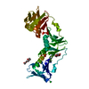
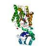
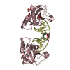
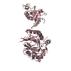
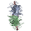
| Title | Crystal Structure of dnaN DNA polymerase III beta subunit from Stenotrophomonas maltophilia K279a | ||||||
 Components Components | DNA polymerase III subunit beta DNA polymerase III holoenzyme DNA polymerase III holoenzyme | ||||||
 Keywords Keywords | DNA BINDING PROTEIN / dnaA / Stenotrophomonas maltophilia / DNA polymerase III beta subunit / Structural Genomics / Seattle Structural Genomics Center for Infectious Disease / SSGCID | ||||||
| Function / homology |  Function and homology informationDNA polymerase III complex / 3'-5' exonuclease activity / DNA replication / DNA-directed DNA polymerase activity / DNA binding / cytoplasm Function and homology informationDNA polymerase III complex / 3'-5' exonuclease activity / DNA replication / DNA-directed DNA polymerase activity / DNA binding / cytoplasmSimilarity search - Function | ||||||
| Biological species |  Stenotrophomonas maltophilia (bacteria) Stenotrophomonas maltophilia (bacteria) | ||||||
| Method | X-RAY DIFFRACTION / SYNCHROTRON / MOLECULAR REPLACEMENT / molecular replacement / Resolution: 2.15 Å | ||||||
 Authors Authors | Seattle Structural Genomics Center for Infectious Disease (SSGCID) | ||||||
| Funding support |  United States, 1items United States, 1items
| ||||||
 Citation Citation | Journal: To Be Published Title: Crystal Structure of dnaN DNA polymerase III beta subunit from Stenotrophomonas maltophilia K279a Authors: Mayclin, S.J. / Lorimer, D.D. / Horanyi, P.S. / Edwards, T.E. | ||||||
| History |
|
- Structure visualization
| Structure viewer | Molecule: MolmilJmol/JSmol |
|---|
- Downloads & links
-Download
| PDBx/mmCIF format | 7rzm.cif.gz | 167.2 KB | Display | PDBx/mmCIF format |
|---|---|---|---|---|
| PDB format | pdb7rzm.ent.gz | 128.9 KB | Display | PDB format |
| PDBx/mmJSON format | 7rzm.json.gz | Tree view | PDBx/mmJSON format | |
| Others |  Other downloads Other downloads |
-Validation report
| Arichive directory | https://data.pdbj.org/pub/pdb/validation_reports/rz/7rzmftp://data.pdbj.org/pub/pdb/validation_reports/rz/7rzm | HTTPS FTP |
|---|
-Related structure data
| Related structure data |  4tszS S: Starting model for refinement |
|---|---|
| Similar structure data |


-Links
 PDBj
PDBj
- Assembly
| Deposited unit | 
| ||||||||
|---|---|---|---|---|---|---|---|---|---|
| 1 |
| ||||||||
| Unit cell |
|
-Components
| #1: Protein | DNA polymerase III holoenzyme / Beta sliding clamp Mass: 41839.316 Da / Num. of mol.: 1 Source method: isolated from a genetically manipulated source Source: (gene. exp.) Stenotrophomonas maltophilia (strain K279a) (bacteria)Strain: K279a / Gene: dnaN, Smlt0002 / Plasmid: StmaA.17987.a.B1 / Production host: Escherichia coli BL21(DE3) (bacteria) / References: UniProt: B2FT80 |
|---|---|
| #2: Water | ChemComp-HOH / Water Mass: 18.015 Da / Num. of mol.: 354 / Source method: isolated from a natural source / Formula: H2O Mass: 18.015 Da / Num. of mol.: 354 / Source method: isolated from a natural source / Formula: H2O |
-Experimental details
-Experiment
| Experiment | Method: X-RAY DIFFRACTION / Number of used crystals: 1 |
|---|
- Sample preparation
| Crystal | Density Matthews: 3.24 Å3/Da / Density % sol: 62 % |
|---|---|
| Crystal grow | Temperature: 287 K / Method: vapor diffusion, sitting drop / pH: 7.5 Details: MORPHEUS F5 (321505f5): 100 mM MOPS/HEPES-Na pH 7.5, 20 mM D-glucose, 20 mM D-mannose, 20 mM D-galactose, 20 mM L-fucose, 20mM D-xylose, 20 mM N-acetyl-D-glucosamine, 10% (w/v) PEG 20000, ...Details: MORPHEUS F5 (321505f5): 100 mM MOPS/HEPES-Na pH 7.5, 20 mM D-glucose, 20 mM D-mannose, 20 mM D-galactose, 20 mM L-fucose, 20mM D-xylose, 20 mM N-acetyl-D-glucosamine, 10% (w/v) PEG 20000, 20% (v/v) PEG MME 550, 16.74 mg/mL StmaA.17987.a.B1.PS38645, direct cryo |
-Data collection
| Diffraction | Mean temperature: 100 K / Serial crystal experiment: N | ||||||||||||||||||||||||||||||||||||||||||||||||||||||||||||||||||||||||||||||||||||||||||||||||||||||||||||||||||||||||||||||||||||||||||||||||||||||||||||||||||||||||||||||||||||||||||||||||||||||||||||||||||
|---|---|---|---|---|---|---|---|---|---|---|---|---|---|---|---|---|---|---|---|---|---|---|---|---|---|---|---|---|---|---|---|---|---|---|---|---|---|---|---|---|---|---|---|---|---|---|---|---|---|---|---|---|---|---|---|---|---|---|---|---|---|---|---|---|---|---|---|---|---|---|---|---|---|---|---|---|---|---|---|---|---|---|---|---|---|---|---|---|---|---|---|---|---|---|---|---|---|---|---|---|---|---|---|---|---|---|---|---|---|---|---|---|---|---|---|---|---|---|---|---|---|---|---|---|---|---|---|---|---|---|---|---|---|---|---|---|---|---|---|---|---|---|---|---|---|---|---|---|---|---|---|---|---|---|---|---|---|---|---|---|---|---|---|---|---|---|---|---|---|---|---|---|---|---|---|---|---|---|---|---|---|---|---|---|---|---|---|---|---|---|---|---|---|---|---|---|---|---|---|---|---|---|---|---|---|---|---|---|---|---|---|
| Diffraction source | Source: SYNCHROTRON / Site: APS / Beamline: 21-ID-F / Wavelength: 0.97872 Å | ||||||||||||||||||||||||||||||||||||||||||||||||||||||||||||||||||||||||||||||||||||||||||||||||||||||||||||||||||||||||||||||||||||||||||||||||||||||||||||||||||||||||||||||||||||||||||||||||||||||||||||||||||
| Detector | Type: RAYONIX MX-300 / Detector: CCD / Date: Jul 15, 2021 / Details: Beryllium Lenses | ||||||||||||||||||||||||||||||||||||||||||||||||||||||||||||||||||||||||||||||||||||||||||||||||||||||||||||||||||||||||||||||||||||||||||||||||||||||||||||||||||||||||||||||||||||||||||||||||||||||||||||||||||
| Radiation | Monochromator: Diamond [111] / Protocol: SINGLE WAVELENGTH / Monochromatic (M) / Laue (L): M / Scattering type: x-ray | ||||||||||||||||||||||||||||||||||||||||||||||||||||||||||||||||||||||||||||||||||||||||||||||||||||||||||||||||||||||||||||||||||||||||||||||||||||||||||||||||||||||||||||||||||||||||||||||||||||||||||||||||||
| Radiation wavelength | Wavelength: 0.97872 Å / Relative weight: 1 | ||||||||||||||||||||||||||||||||||||||||||||||||||||||||||||||||||||||||||||||||||||||||||||||||||||||||||||||||||||||||||||||||||||||||||||||||||||||||||||||||||||||||||||||||||||||||||||||||||||||||||||||||||
| Reflection | Resolution: 2.15→46.47 Å / Num. obs: 29740 / % possible obs: 99.9 % / Redundancy: 5.009 % / Biso Wilson estimate: 35.981 Å2 / CC1/2: 0.999 / Rmerge(I) obs: 0.078 / Rrim(I) all: 0.087 / Χ2: 0.92 / Net I/σ(I): 16.99 / Num. measured all: 148980 | ||||||||||||||||||||||||||||||||||||||||||||||||||||||||||||||||||||||||||||||||||||||||||||||||||||||||||||||||||||||||||||||||||||||||||||||||||||||||||||||||||||||||||||||||||||||||||||||||||||||||||||||||||
| Reflection shell | Diffraction-ID: 1
|
-Phasing
| Phasing | Method: molecular replacement |
|---|
- Processing
| Software |
| |||||||||||||||||||||||||||||||||||||||||||||||||||||||||||||||||||||||||||||||||||||||||||||||||||||||||||||||||||||||||||||
|---|---|---|---|---|---|---|---|---|---|---|---|---|---|---|---|---|---|---|---|---|---|---|---|---|---|---|---|---|---|---|---|---|---|---|---|---|---|---|---|---|---|---|---|---|---|---|---|---|---|---|---|---|---|---|---|---|---|---|---|---|---|---|---|---|---|---|---|---|---|---|---|---|---|---|---|---|---|---|---|---|---|---|---|---|---|---|---|---|---|---|---|---|---|---|---|---|---|---|---|---|---|---|---|---|---|---|---|---|---|---|---|---|---|---|---|---|---|---|---|---|---|---|---|---|---|---|
| Refinement | Method to determine structure: MOLECULAR REPLACEMENT Starting model: 4tsz Resolution: 2.15→46.47 Å / SU ML: 0.23 / Cross valid method: THROUGHOUT / σ(F): 1.35 / Phase error: 22.03 / Stereochemistry target values: ML
| |||||||||||||||||||||||||||||||||||||||||||||||||||||||||||||||||||||||||||||||||||||||||||||||||||||||||||||||||||||||||||||
| Solvent computation | Shrinkage radii: 0.9 Å / VDW probe radii: 1.11 Å / Solvent model: FLAT BULK SOLVENT MODEL | |||||||||||||||||||||||||||||||||||||||||||||||||||||||||||||||||||||||||||||||||||||||||||||||||||||||||||||||||||||||||||||
| Displacement parameters | Biso max: 108.61 Å2 / Biso mean: 35.6982 Å2 / Biso min: 14.5 Å2 | |||||||||||||||||||||||||||||||||||||||||||||||||||||||||||||||||||||||||||||||||||||||||||||||||||||||||||||||||||||||||||||
| Refinement step | Cycle: final / Resolution: 2.15→46.47 Å
| |||||||||||||||||||||||||||||||||||||||||||||||||||||||||||||||||||||||||||||||||||||||||||||||||||||||||||||||||||||||||||||
| LS refinement shell | Refine-ID: X-RAY DIFFRACTION / Rfactor Rfree error: 0 / Total num. of bins used: 15
| |||||||||||||||||||||||||||||||||||||||||||||||||||||||||||||||||||||||||||||||||||||||||||||||||||||||||||||||||||||||||||||
| Refinement TLS params. | Method: refined / Refine-ID: X-RAY DIFFRACTION
| |||||||||||||||||||||||||||||||||||||||||||||||||||||||||||||||||||||||||||||||||||||||||||||||||||||||||||||||||||||||||||||
| Refinement TLS group |
|
