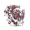
 Movie
Movie Controller


+ Open data

- Basic information
| Entry | Database: PDB / ID: 7rtg | ||||||
|---|---|---|---|---|---|---|---|
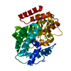
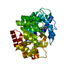
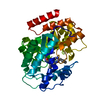
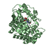
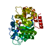
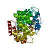
| Title | Crystal Structure of the Human Adenosine Deaminase 1 | ||||||
 Components Components | Adenosine deaminase | ||||||
 Keywords Keywords | HYDROLASE / adenosine deaminase / zinc binding enzyme / holoenzyme | ||||||
| Function / homology |  Function and homology information Function and homology informationpurine nucleotide salvage / Defective ADA disrupts (deoxy)adenosine deamination / mature B cell apoptotic process / xanthine biosynthetic process / negative regulation of penile erection / negative regulation of mucus secretion / penile erection / positive regulation of germinal center formation / negative regulation of adenosine receptor signaling pathway / inosine biosynthetic process ...purine nucleotide salvage / Defective ADA disrupts (deoxy)adenosine deamination / mature B cell apoptotic process / xanthine biosynthetic process / negative regulation of penile erection / negative regulation of mucus secretion / penile erection / positive regulation of germinal center formation / negative regulation of adenosine receptor signaling pathway / inosine biosynthetic process / cytoplasmic vesicle lumen / 2'-deoxyadenosine deaminase activity / amide catabolic process / adenosine deaminase / germinal center B cell differentiation / adenosine catabolic process / purine-containing compound salvage / deaminase activity / adenosine deaminase activity / hypoxanthine salvage / deoxyadenosine catabolic process / dAMP catabolic process / adenosine metabolic process / AMP catabolic process / positive regulation of T cell differentiation in thymus / dATP catabolic process / negative regulation of leukocyte migration / mucus secretion / Ribavirin ADME / regulation of cell-cell adhesion mediated by integrin / response to purine-containing compound / embryonic digestive tract development / allantoin metabolic process / trophectodermal cell differentiation / GMP salvage / Purine salvage / positive regulation of smooth muscle contraction / Peyer's patch development / germinal center formation / negative regulation of mature B cell apoptotic process / AMP salvage / negative regulation of thymocyte apoptotic process / anchoring junction / positive regulation of alpha-beta T cell differentiation / alpha-beta T cell differentiation / positive regulation of heart rate / leukocyte migration / lung alveolus development / positive regulation of T cell receptor signaling pathway / thymocyte apoptotic process / B cell proliferation / smooth muscle contraction / : / positive regulation of calcium-mediated signaling / positive regulation of B cell proliferation / T cell activation / xenobiotic metabolic process / liver development / calcium-mediated signaling / placenta development / negative regulation of inflammatory response / T cell differentiation in thymus / T cell receptor signaling pathway / lysosome / response to hypoxia / cell adhesion / external side of plasma membrane / cell surface / zinc ion binding / membrane / plasma membrane / cytosolSimilarity search - Function | ||||||
| Biological species |  Homo sapiens (human) Homo sapiens (human) | ||||||
| Method | X-RAY DIFFRACTION / SYNCHROTRON / MOLECULAR REPLACEMENT / Resolution: 2.591 Å | ||||||
 Authors Authors | Ma, M.T. / Lieberman, R.L. / Blazeck, J. / Jennings, M.R. | ||||||
| Funding support |  United States, 1items United States, 1items
| ||||||
 Citation Citation | Journal: Acta Crystallogr D Struct Biol / Year: 2022 Title: Catalytically active holo Homo sapiens adenosine deaminase I adopts a closed conformation. Authors: Ma, M.T. / Jennings, M.R. / Blazeck, J. / Lieberman, R.L. | ||||||
| History |
|
- Structure visualization
| Structure viewer | Molecule: MolmilJmol/JSmol |
|---|
- Downloads & links
-Download
| PDBx/mmCIF format | 7rtg.cif.gz | 151.6 KB | Display | PDBx/mmCIF format |
|---|---|---|---|---|
| PDB format | pdb7rtg.ent.gz | 117 KB | Display | PDB format |
| PDBx/mmJSON format | 7rtg.json.gz | Tree view | PDBx/mmJSON format | |
| Others |  Other downloads Other downloads |
-Validation report
| Arichive directory | https://data.pdbj.org/pub/pdb/validation_reports/rt/7rtgftp://data.pdbj.org/pub/pdb/validation_reports/rt/7rtg | HTTPS FTP |
|---|
-Related structure data
| Related structure data | 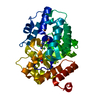 3iarS S: Starting model for refinement |
|---|---|
| Similar structure data |


-Links
 PDBj
PDBj


- Assembly
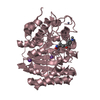
| Deposited unit | 
| |||||||||||||||||||||||||||||||||||||||||||||||||||||||||||||||||||||||
|---|---|---|---|---|---|---|---|---|---|---|---|---|---|---|---|---|---|---|---|---|---|---|---|---|---|---|---|---|---|---|---|---|---|---|---|---|---|---|---|---|---|---|---|---|---|---|---|---|---|---|---|---|---|---|---|---|---|---|---|---|---|---|---|---|---|---|---|---|---|---|---|---|
| 1 | 
| |||||||||||||||||||||||||||||||||||||||||||||||||||||||||||||||||||||||
| 2 | 
| |||||||||||||||||||||||||||||||||||||||||||||||||||||||||||||||||||||||
| Unit cell |
| |||||||||||||||||||||||||||||||||||||||||||||||||||||||||||||||||||||||
| Components on special symmetry positions |
| |||||||||||||||||||||||||||||||||||||||||||||||||||||||||||||||||||||||
| Noncrystallographic symmetry (NCS) | NCS domain:
NCS domain segments: Ens-ID: 1
|
