 Movie
Movie Controller


+ Open data

- Basic information
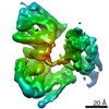
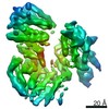
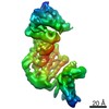
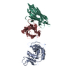
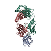
| Entry | Database: PDB / ID: 7rsq | ||||||||||||||||||
|---|---|---|---|---|---|---|---|---|---|---|---|---|---|---|---|---|---|---|---|
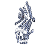
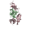
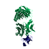
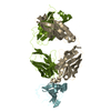
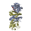
| Title | Cryo-EM structure of KIFBP core | ||||||||||||||||||
 Components Components | KIF-binding protein | ||||||||||||||||||
 Keywords Keywords |  MOTOR PROTEIN / kinesin regulation protein MOTOR PROTEIN / kinesin regulation protein | ||||||||||||||||||
| Function / homology |  Function and homology informationcentral nervous system projection neuron axonogenesis / mitochondrial transport / kinesin binding / neuron projection maintenance / microtubule cytoskeleton organization / in utero embryonic development / cytoskeleton / mitochondrion Function and homology informationcentral nervous system projection neuron axonogenesis / mitochondrial transport / kinesin binding / neuron projection maintenance / microtubule cytoskeleton organization / in utero embryonic development / cytoskeleton / mitochondrionSimilarity search - Function | ||||||||||||||||||
| Biological species |  Homo sapiens (human) Homo sapiens (human) | ||||||||||||||||||
| Method | ELECTRON MICROSCOPY / single particle reconstruction / cryo EM / Resolution: 3.8 Å | ||||||||||||||||||
| Model details | MODEL GENERATED BY ROSETTA VERSION 2020.08+release.cb1caba | ||||||||||||||||||
 Authors Authors | Solon, A.L. / Tan, Z. / Schutt, K.L. / Jepsen, L. / Haynes, S.E. / Nesvizhskii, A.I. / Sept, D. / Stumpff, J. / Ohi, R. / Cianfrocco, M.A. | ||||||||||||||||||
| Funding support |  United States, 5items United States, 5items
| ||||||||||||||||||
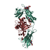
 Citation Citation | Journal: Sci Adv / Year: 2021 Title: Kinesin-binding protein remodels the kinesin motor to prevent microtubule binding. Authors: April L Solon / Zhenyu Tan / Katherine L Schutt / Lauren Jepsen / Sarah E Haynes / Alexey I Nesvizhskii / David Sept / Jason Stumpff / Ryoma Ohi / Michael A Cianfrocco / Abstract: Kinesins are regulated in space and time to ensure activation only in the presence of cargo. Kinesin-binding protein (KIFBP), which is mutated in Goldberg-Shprintzen syndrome, binds to and inhibits ...Kinesins are regulated in space and time to ensure activation only in the presence of cargo. Kinesin-binding protein (KIFBP), which is mutated in Goldberg-Shprintzen syndrome, binds to and inhibits the catalytic motor heads of 8 of 45 kinesin superfamily members, but the mechanism remains poorly defined. Here, we used cryo–electron microscopy and cross-linking mass spectrometry to determine high-resolution structures of KIFBP alone and in complex with two mitotic kinesins, revealing structural remodeling of kinesin by KIFBP. We find that KIFBP remodels kinesin motors and blocks microtubule binding (i) via allosteric changes to kinesin and (ii) by sterically blocking access to the microtubule. We identified two regions of KIFBP necessary for kinesin binding and cellular regulation during mitosis. Together, this work further elucidates the molecular mechanism of KIFBP-mediated kinesin inhibition and supports a model in which structural rearrangement of kinesin motor domains by KIFBP abrogates motor protein activity. | ||||||||||||||||||
| History |
|
- Structure visualization
| Movie |
Movie viewer |
|---|---|
| Structure viewer | Molecule: MolmilJmol/JSmol |
- Downloads & links
-Download
| PDBx/mmCIF format | 7rsq.cif.gz | 94.3 KB | Display | PDBx/mmCIF format |
|---|---|---|---|---|
| PDB format | pdb7rsq.ent.gz | 73.1 KB | Display | PDB format |
| PDBx/mmJSON format | 7rsq.json.gz | Tree view | PDBx/mmJSON format | |
| Others |  Other downloads Other downloads |
-Validation report
| Arichive directory | https://data.pdbj.org/pub/pdb/validation_reports/rs/7rsqftp://data.pdbj.org/pub/pdb/validation_reports/rs/7rsq | HTTPS FTP |
|---|
-Related structure data
| Related structure data | 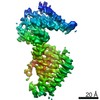 24677MC 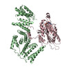 7rsiC 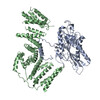 7rypC  7ryqC M: map data used to model this data C: citing same article ( |
|---|---|
| Similar structure data |
-Links
 PDBj
PDBj- Assembly
| Deposited unit | 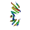
|
|---|---|
| 1 |
|
-Components
| #1: Protein | Mass: 71913.945 Da / Num. of mol.: 1 Source method: isolated from a genetically manipulated source Source: (gene. exp.) Homo sapiens (human) / Gene: KIFBP, KBP, KIAA1279, KIF1BP / Production host:  Escherichia coli BL21 (bacteria) / References: UniProt: Q96EK5 Escherichia coli BL21 (bacteria) / References: UniProt: Q96EK5 |
|---|
-Experimental details
-Experiment
| Experiment | Method: ELECTRON MICROSCOPY |
|---|---|
| EM experiment | Aggregation state: PARTICLE / 3D reconstruction method: single particle reconstruction |
- Sample preparation
| Component | Name: high-resolution structure of KIFBP(core) / Type: COMPLEX / Entity ID: all / Source: RECOMBINANT |
|---|---|
| Molecular weight | Experimental value: NO |
| Source (natural) | Organism: Homo sapiens (human) |
| Source (recombinant) | Organism: Escherichia coli (E. coli) |
| Buffer solution | pH: 7.7 |
| Specimen | Conc.: 4 mg/ml / Embedding applied: NO / Shadowing applied: NO / Staining applied: NO / Vitrification applied: YES |
| Specimen support | Grid type: UltrAuFoil R1.2/1.3 |
| Vitrification | Instrument: FEI VITROBOT MARK IV / Cryogen name: ETHANE |
- Electron microscopy imaging
| Microscopy | Model: TFS GLACIOS |
|---|---|
| Electron gun | Electron source: FIELD EMISSION GUN / Accelerating voltage: 200 kV / Illumination mode: OTHER |
| Electron lens | Mode: BRIGHT FIELDBright-field microscopy |
| Image recording | Electron dose: 60 e/Å2 / Detector mode: COUNTING / Film or detector model: GATAN K2 SUMMIT (4k x 4k) |
- Processing
| EM software |
| |||||||||||||||||||||
|---|---|---|---|---|---|---|---|---|---|---|---|---|---|---|---|---|---|---|---|---|---|---|
| CTF correction | Type: PHASE FLIPPING AND AMPLITUDE CORRECTION | |||||||||||||||||||||
| Particle selection | Num. of particles selected: 128190 | |||||||||||||||||||||
| 3D reconstruction | Resolution: 3.8 Å / Resolution method: FSC 0.143 CUT-OFF / Num. of particles: 128190 / Num. of class averages: 1 / Symmetry type: POINT | |||||||||||||||||||||
| Atomic model building | Protocol: AB INITIO MODEL / Space: REAL |
