 Movie
Movie Controller


+ Open data

- Basic information
| Entry | Database: PDB / ID: 7qoh | ||||||
|---|---|---|---|---|---|---|---|
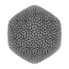
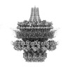
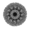
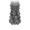
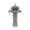
| Title | Unique vertex of the phicrAss001 virion with C5 symmetry imposed | ||||||
 Components Components |
| ||||||
 Keywords Keywords |  VIRUS / crAssphage / bacteriophage / DNA virus / portal / vertex / capsid / connector VIRUS / crAssphage / bacteriophage / DNA virus / portal / vertex / capsid / connector | ||||||
| Function / homology |  Function and homology informationviral capsid, decoration / virion component / viral capsid / symbiont entry into host cell / virion attachment to host cell Function and homology informationviral capsid, decoration / virion component / viral capsid / symbiont entry into host cell / virion attachment to host cellSimilarity search - Function | ||||||
| Biological species |  Bacteroides phage crAss001 (virus) Bacteroides phage crAss001 (virus) | ||||||
| Method | ELECTRON MICROSCOPY / single particle reconstruction / cryo EM / Resolution: 3.32 Å | ||||||
 Authors Authors | Bayfield, O.W. / Shkoporov, A.N. / Yutin, N. / Khokhlova, E.V. / Smith, J.L.R. / Hawkins, D.E.D.P. / Koonin, E.V. / Hill, C. / Antson, A.A. | ||||||
| Funding support |  United Kingdom, 1items United Kingdom, 1items
| ||||||
 Citation Citation | Journal: Nature / Year: 2023 Title: Structural atlas of a human gut crassvirus. Authors: Oliver W Bayfield / Andrey N Shkoporov / Natalya Yutin / Ekaterina V Khokhlova / Jake L R Smith / Dorothy E D P Hawkins / Eugene V Koonin / Colin Hill / Alfred A Antson /   Abstract: CrAssphage and related viruses of the order Crassvirales (hereafter referred to as crassviruses) were originally discovered by cross-assembly of metagenomic sequences. They are the most abundant ...CrAssphage and related viruses of the order Crassvirales (hereafter referred to as crassviruses) were originally discovered by cross-assembly of metagenomic sequences. They are the most abundant viruses in the human gut, are found in the majority of individual gut viromes, and account for up to 95% of the viral sequences in some individuals. Crassviruses are likely to have major roles in shaping the composition and functionality of the human microbiome, but the structures and roles of most of the virally encoded proteins are unknown, with only generic predictions resulting from bioinformatic analyses. Here we present a cryo-electron microscopy reconstruction of Bacteroides intestinalis virus ΦcrAss001, providing the structural basis for the functional assignment of most of its virion proteins. The muzzle protein forms an assembly about 1 MDa in size at the end of the tail and exhibits a previously unknown fold that we designate the 'crass fold', that is likely to serve as a gatekeeper that controls the ejection of cargos. In addition to packing the approximately 103 kb of virus DNA, the ΦcrAss001 virion has extensive storage space for virally encoded cargo proteins in the capsid and, unusually, within the tail. One of the cargo proteins is present in both the capsid and the tail, suggesting a general mechanism for protein ejection, which involves partial unfolding of proteins during their extrusion through the tail. These findings provide a structural basis for understanding the mechanisms of assembly and infection of these highly abundant crassviruses. #1: Journal: Res Sq / Year: 2023Title: Structural atlas of the most abundant human gut virus Authors: Antson, A. / Bayfield, O. / Shkoporov, A. / Yutin, N. / Khokhlova, E. / Smith, J. / Hawkins, D. / Koonin, E. / Hill, C. | ||||||
| History |
|
- Structure visualization
| Structure viewer | Molecule: MolmilJmol/JSmol |
|---|
- Downloads & links
-Download
| PDBx/mmCIF format | 7qoh.cif.gz | 816.1 KB | Display | PDBx/mmCIF format |
|---|---|---|---|---|
| PDB format | pdb7qoh.ent.gz | 673.4 KB | Display | PDB format |
| PDBx/mmJSON format | 7qoh.json.gz | Tree view | PDBx/mmJSON format | |
| Others |  Other downloads Other downloads |
-Validation report
| Arichive directory | https://data.pdbj.org/pub/pdb/validation_reports/qo/7qohftp://data.pdbj.org/pub/pdb/validation_reports/qo/7qoh | HTTPS FTP |
|---|
-Related structure data
| Related structure data |  14090MC 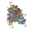 7qofC  7qogC 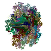 7qoiC  7qojC  7qokC  7qolC M: map data used to model this data C: citing same article ( |
|---|---|
| Similar structure data |


-Links
 PDBj
PDBj- Assembly
| Deposited unit | 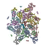
|
|---|---|
| 1 | x 5
|
-Components
-Protein , 5 types, 18 molecules ABCDEFabdefghijklm
| #1: Protein | Mass: 57098.598 Da / Num. of mol.: 6 / Source method: isolated from a natural source / Source: (natural) Bacteroides phage crAss001 (virus) / References: UniProt: A0A385DVU6#2: Protein | Mass: 36154.094 Da / Num. of mol.: 5 / Source method: isolated from a natural source / Source: (natural) Bacteroides phage crAss001 (virus) / References: UniProt: A0A385DVS7#3: Protein | Mass: 11327.080 Da / Num. of mol.: 2 / Source method: isolated from a natural source / Source: (natural) Bacteroides phage crAss001 (virus) / References: UniProt: A0A385DTA3#4: Protein | Mass: 10294.763 Da / Num. of mol.: 3 / Source method: isolated from a natural source / Source: (natural) Bacteroides phage crAss001 (virus) / References: UniProt: A0A385DTC5#5: Protein | Mass: 93122.211 Da / Num. of mol.: 2 / Source method: isolated from a natural source / Source: (natural) Bacteroides phage crAss001 (virus) / References: UniProt: A0A385DT68 |
|---|
-Non-polymers , 1 types, 6 molecules 
| #6: Chemical | ChemComp-MG /  Mass: 24.305 Da / Num. of mol.: 6 / Source method: obtained synthetically / Formula: Mg Mass: 24.305 Da / Num. of mol.: 6 / Source method: obtained synthetically / Formula: Mg |
|---|
-Details
| Has ligand of interest | N |
|---|
-Experimental details
-Experiment
| Experiment | Method: ELECTRON MICROSCOPY |
|---|---|
| EM experiment | Aggregation state: PARTICLE / 3D reconstruction method: single particle reconstruction |
- Sample preparation
| Component | Name: Bacteroides phage crAss001 / Type: VIRUS / Entity ID: #1-#5 / Source: NATURAL |
|---|---|
| Molecular weight | Experimental value: NO |
| Source (natural) | Organism: Bacteroides phage crAss001 (virus) |
| Details of virus | Empty: NO / Enveloped: NO / Isolate: SPECIES / Type: VIRION |
| Buffer solution | pH: 7.5 |
| Specimen | Embedding applied: NO / Shadowing applied: NO / Staining applied: NO / Vitrification applied: YES |
| Vitrification | Cryogen name: ETHANE |
- Electron microscopy imaging
| Experimental equipment |  Model: Titan Krios / Image courtesy: FEI Company |
|---|---|
| Microscopy | Model: FEI TITAN KRIOS |
| Electron gun | Electron source: FIELD EMISSION GUN / Accelerating voltage: 300 kV / Illumination mode: FLOOD BEAM |
| Electron lens | Mode: BRIGHT FIELDBright-field microscopy / Nominal defocus max: 1500 nm / Nominal defocus min: 300 nm |
| Image recording | Electron dose: 51 e/Å2 / Detector mode: INTEGRATING / Film or detector model: FEI FALCON III (4k x 4k) |
- Processing
| Software | Name: PHENIX / Version: 1.19.2_4158: / Classification: refinement | ||||||||||||||||||||||||
|---|---|---|---|---|---|---|---|---|---|---|---|---|---|---|---|---|---|---|---|---|---|---|---|---|---|
| EM software |
| ||||||||||||||||||||||||
| CTF correction | Type: PHASE FLIPPING AND AMPLITUDE CORRECTION | ||||||||||||||||||||||||
| Symmetry | Point symmetry: C5 (5 fold cyclic) | ||||||||||||||||||||||||
| 3D reconstruction | Resolution: 3.32 Å / Resolution method: FSC 0.143 CUT-OFF / Num. of particles: 122709 / Symmetry type: POINT | ||||||||||||||||||||||||
| Atomic model building | B value: 157 / Protocol: AB INITIO MODEL / Space: REAL | ||||||||||||||||||||||||
| Refine LS restraints |
|
