 Movie
Movie Controller


+ Open data

- Basic information
| Entry | Database: PDB / ID: 7pqv | ||||||
|---|---|---|---|---|---|---|---|
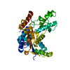
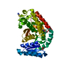
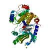
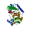
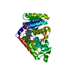
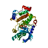
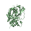
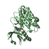
| Title | MEK1 IN COMPLEX WITH COMPOUND 7 | ||||||
 Components Components | Dual specificity mitogen-activated protein kinase kinase 1 | ||||||
 Keywords Keywords |  SIGNALING PROTEIN / MEK1 MITOGEN-ACTIVATED PROTEIN KINASE 1 / KINASE INHIBITOR / ANTI-CANCER DRUG SIGNALING PROTEIN / MEK1 MITOGEN-ACTIVATED PROTEIN KINASE 1 / KINASE INHIBITOR / ANTI-CANCER DRUG | ||||||
| Function / homology |  Function and homology information Function and homology informationepithelial cell proliferation involved in lung morphogenesis / positive regulation of endodermal cell differentiation / placenta blood vessel development / regulation of axon regeneration / mitogen-activated protein kinase kinase / labyrinthine layer development / type B pancreatic cell proliferation / MAP-kinase scaffold activity / cerebellar cortex formation / Signaling by MAP2K mutants ...epithelial cell proliferation involved in lung morphogenesis / positive regulation of endodermal cell differentiation / placenta blood vessel development / regulation of axon regeneration / mitogen-activated protein kinase kinase / labyrinthine layer development / type B pancreatic cell proliferation / MAP-kinase scaffold activity / cerebellar cortex formation / Signaling by MAP2K mutants / regulation of Golgi inheritance / trachea formation / Negative feedback regulation of MAPK pathway / regulation of early endosome to late endosome transport / positive regulation of axonogenesis / regulation of stress-activated MAPK cascade / Frs2-mediated activation / ERBB2-ERBB3 signaling pathway / protein kinase activator activity / endodermal cell differentiation / face development / MAPK3 (ERK1) activation / Bergmann glial cell differentiation / MAP kinase kinase activity / thyroid gland development / Uptake and function of anthrax toxins / Schwann cell development / keratinocyte differentiation / ERK1 and ERK2 cascade / myelination / protein serine/threonine/tyrosine kinase activity / protein serine/threonine kinase activator activity / MAP3K8 (TPL2)-dependent MAPK1/3 activation / insulin-like growth factor receptor signaling pathway / thymus development / Signal transduction by L1 / cell motility / RAF activation / Signaling by high-kinase activity BRAF mutants / MAP2K and MAPK activation / neuron differentiation / positive regulation of protein serine/threonine kinase activity / Signaling by RAF1 mutants / Signaling by moderate kinase activity BRAF mutants / Paradoxical activation of RAF signaling by kinase inactive BRAF / Signaling downstream of RAS mutants / chemotaxis / MAPK cascade / cellular senescence / Signaling by BRAF and RAF1 fusions / late endosome / heart development / scaffold protein binding / protein tyrosine kinase activity / early endosome / positive regulation of ERK1 and ERK2 cascade / protein kinase activity / negative regulation of cell population proliferation / protein phosphorylation / protein serine kinase activity / focal adhesion / protein serine/threonine kinase activity / centrosome / positive regulation of gene expression / positive regulation of DNA-templated transcription / Golgi apparatus / endoplasmic reticulum / signal transduction / mitochondrion / ATP binding / nucleus / plasma membrane / cytosolSimilarity search - Function | ||||||
| Biological species |  Homo sapiens (human) Homo sapiens (human) | ||||||
| Method | X-RAY DIFFRACTION / SYNCHROTRON / MOLECULAR REPLACEMENT / Resolution: 2.13 Å | ||||||
 Authors Authors | Moebitz, H. | ||||||
| Funding support | 1items
| ||||||
 Citation Citation | Journal: J.Med.Chem. / Year: 2022 Title: Discovery of MAP855, an Efficacious and Selective MEK1/2 Inhibitor with an ATP-Competitive Mode of Action. Authors: Poddutoori, R. / Aardalen, K. / Aithal, K. / Barahagar, S.S. / Belliappa, C. / Bock, M. / Chelur, S. / Gerken, A. / Gopinath, S. / Gruenenfelder, B. / Kiffe, M. / Krishnaswami, M. / ...Authors: Poddutoori, R. / Aardalen, K. / Aithal, K. / Barahagar, S.S. / Belliappa, C. / Bock, M. / Chelur, S. / Gerken, A. / Gopinath, S. / Gruenenfelder, B. / Kiffe, M. / Krishnaswami, M. / Langowski, J. / Madapa, S. / Narayanan, K. / Pandit, C. / Panigrahi, S.K. / Perrone, M. / Potakamuri, R.K. / Ramachandra, M. / Ramanathan, A. / Ramos, R. / Sager, E. / Samajdar, S. / Subramanya, H.S. / Thimmasandra, D.S. / Venetsanakos, E. / Mobitz, H. | ||||||
| History |
|
- Structure visualization
| Structure viewer | Molecule: MolmilJmol/JSmol |
|---|
- Downloads & links
-Download
| PDBx/mmCIF format | 7pqv.cif.gz | 80.9 KB | Display | PDBx/mmCIF format |
|---|---|---|---|---|
| PDB format | pdb7pqv.ent.gz | 61.6 KB | Display | PDB format |
| PDBx/mmJSON format | 7pqv.json.gz | Tree view | PDBx/mmJSON format | |
| Others |  Other downloads Other downloads |
-Validation report
| Arichive directory | https://data.pdbj.org/pub/pdb/validation_reports/pq/7pqvftp://data.pdbj.org/pub/pdb/validation_reports/pq/7pqv | HTTPS FTP |
|---|
-Related structure data
| Similar structure data |
|---|


-Links
 PDBj
PDBj


- Assembly
| Deposited unit | 
| ||||||||||||
|---|---|---|---|---|---|---|---|---|---|---|---|---|---|
| 1 |
| ||||||||||||
| Unit cell |
| ||||||||||||
| Components on special symmetry positions |
|
-Components
| #1: Protein | Mass: 38507.414 Da / Num. of mol.: 1 / Fragment: KINASE DOMAIN Source method: isolated from a genetically manipulated source Source: (gene. exp.) Homo sapiens (human) / Gene: MAP2K1, MEK1, PRKMK1 / Production host:  Escherichia coli (E. coli) Escherichia coli (E. coli)References: UniProt: Q02750, mitogen-activated protein kinase kinase | ||||||
|---|---|---|---|---|---|---|---|
| #2: Chemical | ChemComp-80C /   Mass: 410.872 Da / Num. of mol.: 1 / Source method: obtained synthetically / Formula: C22H20ClFN4O / Feature type: SUBJECT OF INVESTIGATION Mass: 410.872 Da / Num. of mol.: 1 / Source method: obtained synthetically / Formula: C22H20ClFN4O / Feature type: SUBJECT OF INVESTIGATION | ||||||
| #3: Chemical |   Mass: 40.078 Da / Num. of mol.: 2 / Source method: obtained synthetically / Formula: Ca Mass: 40.078 Da / Num. of mol.: 2 / Source method: obtained synthetically / Formula: Ca#4: Chemical | ChemComp-MG / |   Mass: 24.305 Da / Num. of mol.: 1 / Source method: obtained synthetically / Formula: Mg Mass: 24.305 Da / Num. of mol.: 1 / Source method: obtained synthetically / Formula: Mg#5: Water | ChemComp-HOH / | Water Mass: 18.015 Da / Num. of mol.: 190 / Source method: isolated from a natural source / Formula: H2O Mass: 18.015 Da / Num. of mol.: 190 / Source method: isolated from a natural source / Formula: H2OHas ligand of interest | Y | |
-Experimental details
-Experiment
| Experiment | Method: X-RAY DIFFRACTION / Number of used crystals: 2 |
|---|
- Sample preparation
| Crystal | Density Matthews: 1.24 Å3/Da / Density % sol: 54 % |
|---|---|
| Crystal grow | Temperature: 310 K / Method: batch mode Details: The purified protein was used in crystallisation trials employing both, a standard screen with approximately 1200 different conditions, as well as crystallisation conditions identified using ...Details: The purified protein was used in crystallisation trials employing both, a standard screen with approximately 1200 different conditions, as well as crystallisation conditions identified using literature data. Condi- tions initially obtained have been optimised using standard strategies, systematically varying parameters critically influencing crystallisation, such as temperature, protein concentration, drop ratio, and others. These conditions were also refined by systematically varying pH or precipitant concentrations. |
-Data collection
| Diffraction | Mean temperature: 100 K / Serial crystal experiment: N |
|---|---|
| Diffraction source | Source: SYNCHROTRON / Site: SLS  / Beamline: X06SA / Wavelength: 0.9798 Å / Beamline: X06SA / Wavelength: 0.9798 Å |
| Detector | Type: DECTRIS PILATUS 6M / Detector: PIXEL / Date: May 2, 2011 |
| Radiation | Protocol: SINGLE WAVELENGTH / Monochromatic (M) / Laue (L): M / Scattering type: x-ray |
| Radiation wavelength | Wavelength: 0.9798 Å / Relative weight: 1 |
| Reflection | Resolution: 2.13→66.82 Å / Num. obs: 22886 / % possible obs: 97.2 % / Redundancy: 5.7 % / Rrim(I) all: 0.081 / Rsym value: 0.075 / Net I/σ(I): 14.66 |
| Reflection shell | Resolution: 2.13→2.3 Å / Redundancy: 5.9 % / Mean I/σ(I) obs: 3.07 / Num. unique obs: 4328 / Rrim(I) all: 0.479 / Rsym value: 0.44 / % possible all: 95.6 |
- Processing
| Software |
| ||||||||||||||||||||||||||||||||||||||||||||||||||||||||||||||||||||||||||||||||||||||||||||||||||||||||||||||
|---|---|---|---|---|---|---|---|---|---|---|---|---|---|---|---|---|---|---|---|---|---|---|---|---|---|---|---|---|---|---|---|---|---|---|---|---|---|---|---|---|---|---|---|---|---|---|---|---|---|---|---|---|---|---|---|---|---|---|---|---|---|---|---|---|---|---|---|---|---|---|---|---|---|---|---|---|---|---|---|---|---|---|---|---|---|---|---|---|---|---|---|---|---|---|---|---|---|---|---|---|---|---|---|---|---|---|---|---|---|---|---|
| Refinement | Method to determine structure: MOLECULAR REPLACEMENT Starting model: NONE Resolution: 2.13→66.82 Å / Cor.coef. Fo:Fc: 0.937 / Cor.coef. Fo:Fc free: 0.905 / SU B: 7.929 / SU ML: 0.195 / Cross valid method: THROUGHOUT / σ(F): 0 / ESU R: 0.259 / ESU R Free: 0.223 / Stereochemistry target values: MAXIMUM LIKELIHOOD / Details: HYDROGENS HAVE BEEN ADDED IN THE RIDING POSITIONS
| ||||||||||||||||||||||||||||||||||||||||||||||||||||||||||||||||||||||||||||||||||||||||||||||||||||||||||||||
| Solvent computation | Ion probe radii: 0.8 Å / Shrinkage radii: 0.8 Å / VDW probe radii: 1.2 Å / Solvent model: BABINET MODEL WITH MASK | ||||||||||||||||||||||||||||||||||||||||||||||||||||||||||||||||||||||||||||||||||||||||||||||||||||||||||||||
| Displacement parameters | Biso max: 81.22 Å2 / Biso mean: 34.619 Å2 / Biso min: 17.76 Å2
| ||||||||||||||||||||||||||||||||||||||||||||||||||||||||||||||||||||||||||||||||||||||||||||||||||||||||||||||
| Refinement step | Cycle: final / Resolution: 2.13→66.82 Å
| ||||||||||||||||||||||||||||||||||||||||||||||||||||||||||||||||||||||||||||||||||||||||||||||||||||||||||||||
| Refine LS restraints |
| ||||||||||||||||||||||||||||||||||||||||||||||||||||||||||||||||||||||||||||||||||||||||||||||||||||||||||||||
| LS refinement shell | Resolution: 2.13→2.185 Å / Rfactor Rfree error: 0 / Total num. of bins used: 20
|
