 Movie
Movie Controller


+ Open data

- Basic information
| Entry | Database: PDB / ID: 3zlx | ||||||
|---|---|---|---|---|---|---|---|
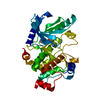
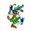
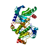
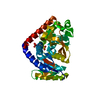
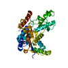
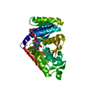
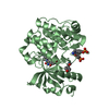
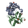
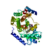
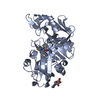
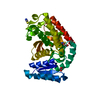
| Title | Crystal structure of MEK1 in complex with fragment 18 | ||||||
 Components Components | DUAL SPECIFICITY MITOGEN-ACTIVATED PROTEIN KINASE KINASE 1 | ||||||
 Keywords Keywords |  TRANSFERASE TRANSFERASE | ||||||
| Function / homology |  Function and homology information Function and homology informationepithelial cell proliferation involved in lung morphogenesis / positive regulation of endodermal cell differentiation / placenta blood vessel development / regulation of axon regeneration / mitogen-activated protein kinase kinase / labyrinthine layer development / type B pancreatic cell proliferation / MAP-kinase scaffold activity / cerebellar cortex formation / Signaling by MAP2K mutants ...epithelial cell proliferation involved in lung morphogenesis / positive regulation of endodermal cell differentiation / placenta blood vessel development / regulation of axon regeneration / mitogen-activated protein kinase kinase / labyrinthine layer development / type B pancreatic cell proliferation / MAP-kinase scaffold activity / cerebellar cortex formation / Signaling by MAP2K mutants / regulation of Golgi inheritance / trachea formation / Negative feedback regulation of MAPK pathway / regulation of early endosome to late endosome transport / positive regulation of axonogenesis / regulation of stress-activated MAPK cascade / Frs2-mediated activation / ERBB2-ERBB3 signaling pathway / protein kinase activator activity / endodermal cell differentiation / face development / MAPK3 (ERK1) activation / Bergmann glial cell differentiation / MAP kinase kinase activity / thyroid gland development / Uptake and function of anthrax toxins / Schwann cell development / keratinocyte differentiation / ERK1 and ERK2 cascade / myelination / protein serine/threonine/tyrosine kinase activity / protein serine/threonine kinase activator activity / MAP3K8 (TPL2)-dependent MAPK1/3 activation / insulin-like growth factor receptor signaling pathway / thymus development / Signal transduction by L1 / cell motility / RAF activation / Signaling by high-kinase activity BRAF mutants / MAP2K and MAPK activation / neuron differentiation / positive regulation of protein serine/threonine kinase activity / Signaling by RAF1 mutants / Signaling by moderate kinase activity BRAF mutants / Paradoxical activation of RAF signaling by kinase inactive BRAF / Signaling downstream of RAS mutants / chemotaxis / MAPK cascade / cellular senescence / Signaling by BRAF and RAF1 fusions / late endosome / heart development / scaffold protein binding / protein tyrosine kinase activity / early endosome / positive regulation of ERK1 and ERK2 cascade / protein kinase activity / negative regulation of cell population proliferation / protein phosphorylation / protein serine kinase activity / focal adhesion / protein serine/threonine kinase activity / centrosome / positive regulation of gene expression / positive regulation of DNA-templated transcription / Golgi apparatus / endoplasmic reticulum / signal transduction / mitochondrion / ATP binding / nucleus / plasma membrane / cytosolSimilarity search - Function | ||||||
| Biological species |  HOMO SAPIENS (human) HOMO SAPIENS (human) | ||||||
| Method | X-RAY DIFFRACTION / SYNCHROTRON / MOLECULAR REPLACEMENT / Resolution: 2.2 Å | ||||||
 Authors Authors | Amaning, K. / Lowinsky, M. / Vallee, F. / Steier, V. / Marcireau, C. / Ugolini, A. / Delorme, C. / McCort, G. / Andouche, C. / Vougier, S. ...Amaning, K. / Lowinsky, M. / Vallee, F. / Steier, V. / Marcireau, C. / Ugolini, A. / Delorme, C. / McCort, G. / Andouche, C. / Vougier, S. / Llopart, S. / Halland, N. / Rak, A. | ||||||
 Citation Citation | Journal: Bioorg.Med.Chem.Lett. / Year: 2013 Title: The Use of Virtual Screening and Differential Scanning Fluorimetry for the Rapid Identification of Fragments Active Against Mek1. Authors: Amaning, K. / Lowinski, M. / Vallee, F. / Steier, V. / Marcireau, C. / Ugolini, A. / Delorme, C. / Foucalt, F. / Mccort, G. / Derimay, N. / Andouche, C. / Vougier, S. / Llopart, S. / Halland, N. / Rak, A. | ||||||
| History |
|
- Structure visualization
| Structure viewer | Molecule: MolmilJmol/JSmol |
|---|
- Downloads & links
-Download
| PDBx/mmCIF format | 3zlx.cif.gz | 77.5 KB | Display | PDBx/mmCIF format |
|---|---|---|---|---|
| PDB format | pdb3zlx.ent.gz | 57.5 KB | Display | PDB format |
| PDBx/mmJSON format | 3zlx.json.gz | Tree view | PDBx/mmJSON format | |
| Others |  Other downloads Other downloads |
-Validation report
| Arichive directory | https://data.pdbj.org/pub/pdb/validation_reports/zl/3zlxftp://data.pdbj.org/pub/pdb/validation_reports/zl/3zlx | HTTPS FTP |
|---|
-Related structure data


-Links
 PDBj
PDBj


- Assembly
| Deposited unit | 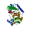
| ||||||||
|---|---|---|---|---|---|---|---|---|---|
| 1 |
| ||||||||
| Unit cell |
| ||||||||
| Components on special symmetry positions |
|
-Components
| #1: Protein | Mass: 38916.879 Da / Num. of mol.: 1 / Mutation: YES Source method: isolated from a genetically manipulated source Source: (gene. exp.) HOMO SAPIENS (human) / Cell line (production host): Sf21 / Production host:   SPODOPTERA FRUGIPERDA (fall armyworm) SPODOPTERA FRUGIPERDA (fall armyworm)References: UniProt: Q02750, mitogen-activated protein kinase kinase |
|---|---|
| #2: Chemical | ChemComp-5EZ /   Mass: 278.734 Da / Num. of mol.: 1 / Source method: obtained synthetically / Formula: C14H15ClN2O2 Mass: 278.734 Da / Num. of mol.: 1 / Source method: obtained synthetically / Formula: C14H15ClN2O2 |
| #3: Water | ChemComp-HOH / Water Mass: 18.015 Da / Num. of mol.: 91 / Source method: isolated from a natural source / Formula: H2O Mass: 18.015 Da / Num. of mol.: 91 / Source method: isolated from a natural source / Formula: H2O |
-Experimental details
-Experiment
| Experiment | Method: X-RAY DIFFRACTION / Number of used crystals: 1 |
|---|
- Sample preparation
| Crystal | Density Matthews: 2.43 Å3/Da / Density % sol: 49.46 % / Description: NONE |
|---|---|
| Crystal grow | pH: 7.7 Details: Appropriate single crystals were grown using this set-up in PEG 4000 18%, Tris 100mM pH7.7, DMSO 2% and CaCl2 0.2M and were extracted from the low volume drops, cryo-protected in mother ...Details: Appropriate single crystals were grown using this set-up in PEG 4000 18%, Tris 100mM pH7.7, DMSO 2% and CaCl2 0.2M and were extracted from the low volume drops, cryo-protected in mother liquor with 20% glycerol and flash cooled for synchrotron collection |
-Data collection
| Diffraction | Mean temperature: 100 K |
|---|---|
| Diffraction source | Source: SYNCHROTRON / Site: ESRF  / Beamline: ID23-1 / Type: ESRF / Wavelength: 0.9795 / Beamline: ID23-1 / Type: ESRF / Wavelength: 0.9795 |
| Detector | Date: Jun 7, 2011 |
| Radiation | Protocol: SINGLE WAVELENGTH / Monochromatic (M) / Laue (L): M / Scattering type: x-ray |
| Radiation wavelength | Wavelength: 0.9795 Å / Relative weight: 1 |
| Reflection | Resolution: 2.2→63.81 Å / Num. obs: 20598 / % possible obs: 99.7 % / Observed criterion σ(I): 2 / Redundancy: 6.2 % / Biso Wilson estimate: 40.77 Å2 / Rmerge(I) obs: 0.08 / Net I/σ(I): 16.3 |
| Reflection shell | Resolution: 2.2→2.32 Å / Redundancy: 6.4 % / Rmerge(I) obs: 0.47 / Mean I/σ(I) obs: 6 / % possible all: 99.8 |
- Processing
| Software |
| ||||||||||||||||||||||||||||||||||||||||||||||||||||||||||||||||||||||||||||||||||||||||||||||||||||||||||||||||||
|---|---|---|---|---|---|---|---|---|---|---|---|---|---|---|---|---|---|---|---|---|---|---|---|---|---|---|---|---|---|---|---|---|---|---|---|---|---|---|---|---|---|---|---|---|---|---|---|---|---|---|---|---|---|---|---|---|---|---|---|---|---|---|---|---|---|---|---|---|---|---|---|---|---|---|---|---|---|---|---|---|---|---|---|---|---|---|---|---|---|---|---|---|---|---|---|---|---|---|---|---|---|---|---|---|---|---|---|---|---|---|---|---|---|---|---|
| Refinement | Method to determine structure: MOLECULAR REPLACEMENT / Resolution: 2.2→49.49 Å / Cor.coef. Fo:Fc: 0.937 / Cor.coef. Fo:Fc free: 0.925 / SU R Cruickshank DPI: 0.221 / Cross valid method: THROUGHOUT / σ(F): 0 / SU R Blow DPI: 0.236 / SU Rfree Blow DPI: 0.187 / SU Rfree Cruickshank DPI: 0.182
| ||||||||||||||||||||||||||||||||||||||||||||||||||||||||||||||||||||||||||||||||||||||||||||||||||||||||||||||||||
| Displacement parameters | Biso mean: 49.59 Å2
| ||||||||||||||||||||||||||||||||||||||||||||||||||||||||||||||||||||||||||||||||||||||||||||||||||||||||||||||||||
| Refine analyze | Luzzati coordinate error obs: 0.273 Å | ||||||||||||||||||||||||||||||||||||||||||||||||||||||||||||||||||||||||||||||||||||||||||||||||||||||||||||||||||
| Refinement step | Cycle: LAST / Resolution: 2.2→49.49 Å
| ||||||||||||||||||||||||||||||||||||||||||||||||||||||||||||||||||||||||||||||||||||||||||||||||||||||||||||||||||
| Refine LS restraints |
| ||||||||||||||||||||||||||||||||||||||||||||||||||||||||||||||||||||||||||||||||||||||||||||||||||||||||||||||||||
| LS refinement shell | Resolution: 2.2→2.32 Å / Total num. of bins used: 10
|
